The Rabbit anti GFP-Tag Polyconal Antibody (CABE011) is a high-quality antibody developed for reliable detection and analysis of target proteins. The green fluorescent protein (GFP) is a protein composed of 238 amino acid residues (26.9 kDa) that exhibits bright green fluorescence when exposed to light in the blue to ultraviolet range. Although many other marine organisms have similar green fluorescent proteins, GFP traditionally refers to the protein first isolated from the jellyfish Aequorea victoria. The GFP from A. victoria has a major excitation peak at a wavelength of 395 nm and a minor one at 475 nm. Its emission peak is at 509 nm, which is in the lower green portion of the visible spectrum. The GFP from the sea pansy (Renilla reniformis) has a single major excitation peak at 498 nm. GFP makes for an excellent tool in many forms of biology due to its ability to form internal chromophore without requiring any accessory cofactors, gene products, or enzymes / substrates other than molecular oxygen.In cell and molecular biology, the GFP gene is frequently used as a reporter of expression. It has been used in modified forms to make biosensors, and many animals have been created that express GFP, which demonstrates a proof of concept that a gene can be expressed throughout a given organism, in selected organs, or in cells of interest. GFP can be introduced into animals or other species through transgenic techniques, and maintained in their genome and that of their offspring. To date, GFP has been expressed in many species, including bacteria, yeasts, fungi, fish and mammals, including in human cells. RRID AB_2771922 Gene ID Swiss Prot Synonym GFP;GFP tag;GFP-tag
This antibody is validated for use in WB, IF/ICC applications and has demonstrated reactivity against Species independent samples.
Product Name:
Rabbit anti GFP-Tag Polyconal Antibody
SKU:
CABE011
Size:
100μL, 200μL, 50μL
Reactivity:
Species independent
Conjugate:
Unconjugated
Immunogen:
Recombinant protein (or fragment).This information is considered to be commercially sensitive.
Tested Applications:
WBIF/ICC
Recommended Dilution:
WB
1:2000 - 1:20000
IF/ICC
1:50 - 1:200
Synonyms:
GFP, GFP tag, GFP-tag
Positive Sample:
293T, insect
Calculated MW:
27kDa
Observed MW:
25kDa
The green fluorescent protein (GFP) is a protein composed of 238 amino acid residues (26.9 kDa) that exhibits bright green fluorescence when exposed to light in the blue to ultraviolet range. Although many other marine organisms have similar green fluorescent proteins, GFP traditionally refers to the protein first isolated from the jellyfish Aequorea victoria. The GFP from A. victoria has a major excitation peak at a wavelength of 395 nm and a minor one at 475 nm. Its emission peak is at 509 nm, which is in the lower green portion of the visible spectrum. The GFP from the sea pansy (Renilla reniformis) has a single major excitation peak at 498 nm. GFP makes for an excellent tool in many forms of biology due to its ability to form internal chromophore without requiring any accessory cofactors, gene products, or enzymes / substrates other than molecular oxygen.In cell and molecular biology, the GFP gene is frequently used as a reporter of expression. It has been used in modified forms to make biosensors, and many animals have been created that express GFP, which demonstrates a proof of concept that a gene can be expressed throughout a given organism, in selected organs, or in cells of interest. GFP can be introduced into animals or other species through transgenic techniques, and maintained in their genome and that of their offspring. To date, GFP has been expressed in many species, including bacteria, yeasts, fungi, fish and mammals, including in human cells. RRID AB_2771922 Gene ID Swiss Prot Synonym GFP;GFP tag;GFP-tag
Purification Method
Affinity purification
RRID
AB_2771922
Buffer Information
Store at -20℃. Avoid freeze / thaw cycles. Buffer: PBS containing 50% glycerol, preserved with proclin300 or sodium azide, pH 7.3.
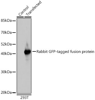
Western blot analysis of extracts of normal 293T cells and 293T transfected with GFP-tagged fusion protein,using Rabbit anti GFP-Tag pAb (CABE011) at 1:1000 dilution. Secondary antibody: HRP-conjugated Goat anti-Rabbit IgG (H+L) (AS014) at 1:10000 dilution. Lysates/proteins: 25μg per lane. Blocking buffer: 3% nonfat dry milk in TBST. Detection: ECL Basic Kit (AbGn00020). Exposure time: 10s.
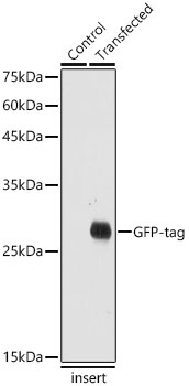
Western blot analysis of extracts of normal Eukaryotic expression of GFP and Eukaryotic expression of GFP transfected with GFP Protein, using Rabbit anti GFP-Tag pAb (CABE011) at 1:20000 dilution. Secondary antibody: HRP-conjugated Goat anti-Rabbit IgG (H+L) (AS014) at 1:10000 dilution. Lysates/proteins: 25μg per lane. Blocking buffer: 3% nonfat dry milk in TBST. Detection: ECL Basic Kit (AbGn00020). Exposure time: 1s.
")
")
")
")
")
")
")