Description
| Product Name: | Rat GDH/GLDH (Glutamate Dehydrogenase) CLIA Kit |
| SKU: | AEES03371 |
| Size: | 96 Assays |
| Detection Method: | Chemiluminescence |
| Assay Type: | Sandwich-ELISA |
| Sensitivity: | 75 pg/mL |
| Range: | 125-8000 pg/mL |
| Reactivity: | Rat |
| Sample Volume: | 100 μL |
| Total Assay Time: | 4 h 30 min |
| Recovery: | 80%-120% |
| Kit Components: |
| |||||||||||||||||||||||||||||||||||||||||||||||||||||||
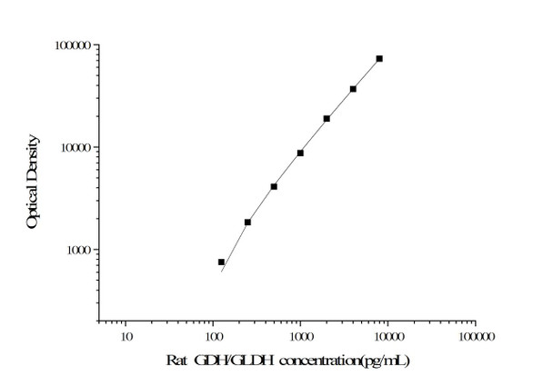
This CLIA kit uses the Sandwich-CLIA principle. The micro CLIA plate provided in this kit has been pre-coated with an antibody specific to Rat GDH/GLDH. Samples or Standards are added to the micro CLIA plate wells and combined with the specific antibody. Then a biotinylated detection antibody specific for Rat GDH/GLDH and Avidin-Horseradish Peroxidase (HRP) conjugate are added successively to each micro plate well and incubated. Free components are washed away. The substrate solution is added to each well. Only those wells that contain Rat GDH/GLDH, biotinylated detection antibody and Avidin-HRP conjugate will appear luminescence. The Relative light unit (RLU) value is measured by the Chemiluminescence immunoassay analyzer at 425nm. The RLU value is positively associated with the concentration of Rat GDH/GLDH. You can calculate the concentration of Rat GDH/GLDH in the samples by comparing the RLU value of the samples to the standard curve.
| Standard: |
| ||||||||||||||||||||||||||||||||||||||||||
| Linearity: |
| ||||||||||||||||||||||||||||||||||||||||||
| Recovery: |
| ||||||||||||||||||||||||||||||||||||||||||
| Precision: |
| ||||||||||||||||||||||||||||||||||||||||||
*Note:The below protocol is a sample protocol. Protocols are specific to each batch/lot. For the correct instructions please follow the protocol included in your kit.
| Step | Protocol |
| 1. | Set standard, test sample and control (zero) wells on the pre-coated plate and record their positions. It is recommended to measure each standard and sample in duplicate. Note: add all solutions to the bottom of the plate wells while avoiding contact with the well walls. Ensure solutions do not foam when adding to the wells. |
| 2. | Aliquot 100µL of standard solutions into the standard wells. |
| 3. | Add 100µL of Sample / Standard dilution buffer into the control (zero) well. |
| 4. | Add 100µL of properly diluted sample (serum, plasma, tissue homogenates and other biological fluids) into test sample wells. |
| 5. | Cover the plate with the sealer provided in the kit and incubate for 90 min at 37°C. Note: add all solutions to the bottom of the plate wells while avoiding contact with the well walls. Ensure solutions do not foam when adding to the wells. |
| 6. | Aspirate the liquid from each well, do not wash. Immediately add 100µL of Biotinylated Detection Ab working solution to each well. Cover the plate with a plate seal and gently mix. Incubate for 1 hour at 37°C. |
| 7. | Aspirate or decant the solution from the plate and add 350µL of wash buffer to each well and incubate for 1-2 minutes at room temperature. Aspirate the solution from each well and clap the plate on absorbent filter paper to dry. Repeat this process 3 times. Note: a microplate washer can be used in this step and other wash steps. |
| 8. | Add 100µL of HRP Conjugate working solution to each well. Cover with a plate seal and incubate for 30 min at 37°C. |
| 9. | Aspirate or decant the solution from each well. Repeat the wash process five times as conducted in step 7. |
| 10. | Add 100µL of Substrate mixture solution to each well. Cover with a new plate seal and incubate for no more than 5 min at 37°C. Protect the plate from light. |
| 11. | Determine the RLU value of each well immediately. |
When carrying out an ELISA assay it is important to prepare your samples in order to achieve the best possible results. Below we have a list of procedures for the preparation of samples for different sample types.
| Sample Type | Protocol |
| Serum | Allow samples to clot for 2 hours at room temperature or overnight at 4°C before centrifugation for 15 min at 1000×g at 2~8°C. Collect the supernatant to carry out the assay. Blood collection tubes should be disposable and endotoxin-free. |
| Plasma | Collect plasma using EDTA or heparin as an anticoagulant. Centrifuge samples for 15 min at 1000×g at 2~8°C within 30 min of collection. Collect the supernatant to carry out the assay. Hemolysed samples are not suitable for ELISA assay. |
| Cell lysates | For adherent cells, gently wash the cells with pre-cooled PBS and dissociate the cells using trypsin. Collect the cell suspension in a tube and centrifuge for 5 min at 1000×g. Discard the medium and wash the cells 3 times with precooled PBS. For each 1×10⁶ cells, add 150-250µL of pre-cooled PBS to keep the cells suspended. Optimal cell concentration is 1 million/ml. To release cellular components, dilute the cell pellet in PBS and use 3-4 freeze-thaw cycles in liquid Nitrogen (commercial lysis buffers can be used according to manufacturer’s instructions). Centrifuge at 4°C for 20 mins at 2000-3000 rpm to pellet debris and remove clear supernatant to clean microcentrifuge tube for ELISA or storage. |
| Tissue homogenates | It is recommended to get detailed references from the literature before analyzing different tissue types. For general information, hemolysed blood may affect the results, so the tissues should be minced into small pieces and rinsed in ice-cold PBS (0.01M, pH=7.4) to remove excess blood thoroughly. Tissue pieces should be weighed and then homogenized in PBS (tissue weight (g): PBS (mL) volume=1:9) with a glass homogenizer on ice. To further break down the cells, you can sonicate the suspension with an ultrasonic cell disrupter or subject it to freeze-thaw cycles. The homogenates are then centrifuged for 5 min at 5000×g to get the supernatant. |
| Cell culture supernatant or other biological fluids | Centrifuge samples for 20 min at 1000×g at 2~8°C. Collect the supernatant to carry out the assay. |
| Sample Type: | Serum, plasma and other biological fluids |
| Specificity: | This kit recognizes Rat GDH/GLDH in samples.No significant cross-reactivity or interference between Rat GDH/GLDH and analogues was observed |
| Storage: | 2-8℃/-20℃ |
| Expiration Date: | 6 months |


