The ROCK1 Polyclonal Antibody (CAB0144) is a high-quality antibody developed for reliable detection and analysis of target proteins. This antibody, produced in rabbits, is highly specific and validated for use in Western blot applications with human samples.ROCK1 is a serine/threonine kinase that is known to be involved in various cellular processes, including actin cytoskeleton organization and cell shape changes. Dysregulation of the ROCK1 pathway has been implicated in diseases such as cancer, cardiovascular disorders, and neurodegenerative diseases, making it an important target for therapeutic intervention.
This antibody is validated for use in WB, IP, ELISA, IF-P applications and has demonstrated reactivity against Human, Mouse, Rat samples.
Product Name:
ROCK1 Polyclonal Antibody
SKU:
CAB0144
Size:
20μL, 100μL
Reactivity:
Human, Mouse, Rat
Conjugate:
Unconjugated
Immunogen:
Recombinant protein (or fragment).This information is considered to be commercially sensitive.
This gene encodes a protein serine/threonine kinase that is activated when bound to the GTP-bound form of Rho. The small GTPase Rho regulates formation of focal adhesions and stress fibers of fibroblasts, as well as adhesion and aggregation of platelets and lymphocytes by shuttling between the inactive GDP-bound form and the active GTP-bound form. Rho is also essential in cytokinesis and plays a role in transcriptional activation by serum response factor. This protein, a downstream effector of Rho, phosphorylates and activates LIM kinase, which in turn, phosphorylates cofilin, inhibiting its actin-depolymerizing activity. A pseudogene, related to this gene, is also located on chromosome 18.
Purification Method
Affinity purification
Gene ID
6093
RRID
AB_2772058
Buffer Information
Store at -20℃. Avoid freeze / thaw cycles. Buffer: PBS containing 50% glycerol, preserved with proclin300 or sodium azide, pH 7.3.
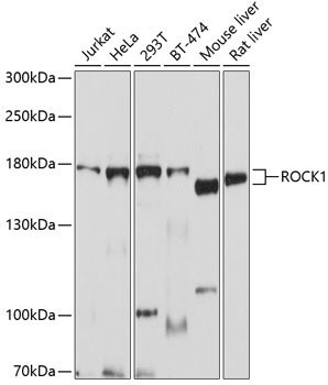
Western blot analysis of various lysates using ROCK1 Rabbit pAb (CAB0144) at 1:1000 dilution. Secondary antibody: HRP-conjugated Goat anti-Rabbit IgG (H+L) (CABS014) at 1:10000 dilution. Lysates/proteins: 25μg per lane. Blocking buffer: 3% nonfat dry milk in TBST. Detection: ECL Enhanced Kit (AbGn00021). Exposure time: 10s.
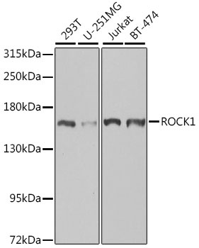
Western blot analysis of various lysates using ROCK1 Rabbit pAb (CAB0144) at 1:1000 dilution. Secondary antibody: HRP-conjugated Goat anti-Rabbit IgG (H+L) (CABS014) at 1:10000 dilution. Lysates/proteins: 25μg per lane. Blocking buffer: 3% nonfat dry milk in TBST. Detection: ECL Basic Kit (AbGn00020). Exposure time: 90s.
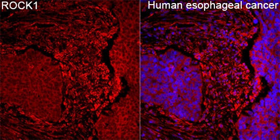
Immunofluorescence analysis of Human esophageal cancer tissue using ROCK1 Rabbit pAb (CAB0144) at a dilution of 1:100 (40x lens). Secondary antibody: Cy3-conjugated Goat anti-Rabbit IgG (H+L)(CABS007) at 1:500 dilution. Blue: DAPI for nuclear staining. High pressure antigen retrieval performed with 0.01M Citrate Buffer (pH 6.0) prior to IF staining.
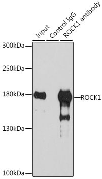
Immunoprecipitation analysis of 200 μg extracts of 293T cells, using 3 μg ROCK1 antibody (CAB0144). Western blot was performed from the immunoprecipitate using ROCK1 antibody (CAB0144) at a dilution of 1:1000.
")
")


")
 at 1:10000 dilution. Lysates/proteins: 25ug per lane. Blocking buffer: 3% nonfat dry milk in TBST. Detection: ECL Basic Kit. Exposure time: 1s.")