The SDC2 Antibody (CAB1810) is a high-quality antibody developed for reliable detection and analysis of target proteins. This antibody, produced in rabbits, shows high reactivity with human samples and is validated for use in various applications, including Western blotting.Syndecan-2 plays a crucial role in cell-extracellular matrix interactions, cell proliferation, and tissue repair processes. Its dysregulation has been linked to various pathological conditions, including cancer, inflammation, and fibrosis.
This antibody is validated for use in WB, IHC-P, ELISA applications and has demonstrated reactivity against Human, Mouse, Rat samples.
Product Name:
SDC2 Antibody
SKU:
CAB1810
Size:
20μL, 100μL
Reactivity:
Human, Mouse, Rat
Conjugate:
Unconjugated
Immunogen:
Recombinant protein (or fragment).This information is considered to be commercially sensitive.
Recommended starting concentration is 1 μg/mL. Please optimize the concentration based on your specific assay requirements.
Synonyms:
HSPG, CD362, HSPG1, SYND2, SDC2
Positive Sample:
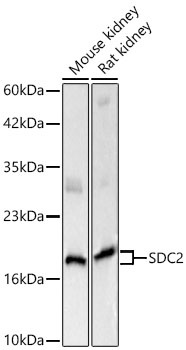
Mouse kidney, Rat kidney
Cellular Localization:
Membrane, Single-Pass Type I Membrane Protein.
Calculated MW:
22kDa
Observed MW:
50kDa/22kDa
The protein encoded by this gene is a transmembrane (type I) heparan sulfate proteoglycan and is a member of the syndecan proteoglycan family. The syndecans mediate cell binding, cell signaling, and cytoskeletal organization and syndecan receptors are required for internalization of the HIV-1 tat protein. The syndecan-2 protein functions as an integral membrane protein and participates in cell proliferation, cell migration and cell-matrix interactions via its receptor for extracellular matrix proteins. Altered syndecan-2 expression has been detected in several different tumor types.
Purification Method
Affinity purification
Gene ID
6383
RRID
AB_2763848
Buffer Information
Store at -20℃. Avoid freeze / thaw cycles. Buffer: PBS with 0.09% sodium azide,50% glycerol,pH7.3.
Western blot analysis of various lysates using SDC2 Rabbit pAb (CAB1810) at 1:1000 dilution. Secondary antibody: HRP-conjugated Goat anti-Rabbit IgG (H+L) (CABS014) at 1:10000 dilution. Lysates / proteins: 25 μg per lane. Blocking buffer: 3 % nonfat dry milk in TBST. Detection: ECL Basic Kit (AbGn00020). Exposure time: 5s.
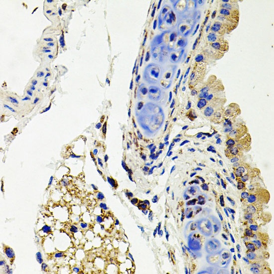
Immunohistochemistry analysis of paraffin-embedded Mouse lung using SDC2 Rabbit pAb (CAB1810) at dilution of 1:100 (40x lens). Microwave antigen retrieval performed with 0.01M PBS Buffer (pH 7.2) prior to IHC staining.
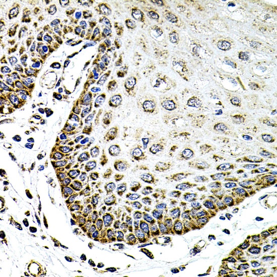
Immunohistochemistry analysis of paraffin-embedded Human esophagus using SDC2 Rabbit pAb (CAB1810) at dilution of 1:100 (40x lens). Microwave antigen retrieval performed with 0.01M PBS Buffer (pH 7.2) prior to IHC staining.