The HSP47/SERPINH1 Polyclonal Antibody (CAB13474) is a high-quality antibody developed for reliable detection and analysis of target proteins. This antibody, generated in rabbits, is highly specific to human samples and has been validated for use in Western blot applications.SERPINH1, also known as heat shock protein 47 (HSP47), plays a crucial role in collagen biosynthesis by assisting in the proper folding and stabilization of collagen triple helices. Dysregulation of SERPINH1 has been implicated in various pathological conditions, including fibrosis, connective tissue disorders, and cancer.
This antibody is validated for use in WB, IF/ICC, ELISA applications and has demonstrated reactivity against Human, Mouse samples.
Product Name:
HSP47/SERPINH1 Polyclonal Antibody
SKU:
CAB13474
Size:
20μL, 100μL
Reactivity:
Human, Mouse
Conjugate:
Unconjugated
Immunogen:
Recombinant protein (or fragment).This information is considered to be commercially sensitive.
This gene encodes a member of the serpin superfamily of serine proteinase inhibitors. The encoded protein is localized to the endoplasmic reticulum and plays a role in collagen biosynthesis as a collagen-specific molecular chaperone. Autoantibodies to the encoded protein have been found in patients with rheumatoid arthritis. Expression of this gene may be a marker for cancer, and nucleotide polymorphisms in this gene may be associated with preterm birth caused by preterm premature rupture of membranes. Alternatively spliced transcript variants have been observed for this gene, and a pseudogene of this gene is located on the short arm of chromosome 9.
Purification Method
Affinity purification
Gene ID
871
RRID
AB_2760336
Buffer Information
Store at -20℃. Avoid freeze / thaw cycles. Buffer: PBS with 0.09% Sodium azide,50% glycerol,pH7.3.
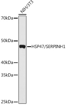
Western blot analysis of various lysates using HSP47/SERPINH1 Rabbit pAb (CAB13474) at 1:1000 dilution. Secondary antibody: HRP-conjugated Goat anti-Rabbit IgG (H+L) (CABS014) at 1:10000 dilution. Lysates/proteins: 25μg per lane. Blocking buffer: 3% nonfat dry milk in TBST. Detection: ECL Basic Kit (AbGn00020). Exposure time: 180s.
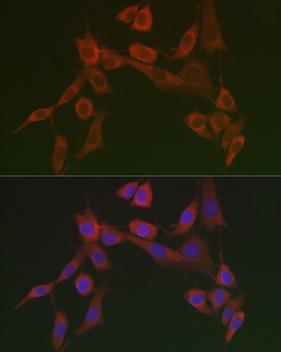
Immunofluorescence analysis of NIH/3T3 cells using HSP47/SERPINH1 Rabbit pAb (CAB13474) at dilution of 1:100 (40x lens). Secondary antibody: Cy3-conjugated Goat anti-Rabbit IgG (H+L) (CABS007) at 1:500 dilution. Blue: DAPI for nuclear staining.


")

 at 1:10000 dilution. Lysates/proteins: 25ug per lane. Blocking buffer: 3% nonfat dry milk in TBST. Detection: ECL Enhanced Kit. Exposure time: 300s.")