The SFPQ Monoclonal Antibody (CAB3494) is a high-quality antibody developed for reliable detection and analysis of target proteins. This antibody, raised in rabbits, is highly reactive with human samples and has been validated for use in various applications, including Western blot and immunohistochemistry.The SFPQ protein plays crucial roles in regulating RNA splicing, DNA repair, and gene expression, making it a key player in cellular processes such as development, differentiation, and stress response. The SFPQ Rabbit Monoclonal Antibody allows for the detection and analysis of SFPQ protein in different cell types, providing researchers with a valuable tool for studying the molecular mechanisms underlying these processes.
This antibody is validated for use in WB, IHC-P, IF/ICC, IP, ELISA applications and has demonstrated reactivity against Human, Mouse, Rat samples.
Product Name:
SFPQ Monoclonal Antibody
SKU:
CAB3494
Size:
20μL, 100μL
Reactivity:
Human, Mouse, Rat
Clone Number:
ARC0788
Conjugate:
Unconjugated
Immunogen:
Synthetic peptide. This information is considered to be commercially sensitive.
0.5μg-4μg antibody for 200μg-400μg extracts of whole cells
ELISA
Recommended starting concentration is 1 μg/mL. Please optimize the concentration based on your specific assay requirements.
Synonyms:
PSF, POMP100, PPP1R140, SFPQ
Positive Sample:
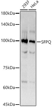
293F, HeLa
Cellular Localization:
Cytoplasm, Nucleus Matrix, Nucleus Speckle.
Calculated MW:
76kDa
Observed MW:
100kDa
Enables DNA binding activity; histone deacetylase binding activity; and protein homodimerization activity. Involved in several processes, including alternative mRNA splicing, via spliceosome; positive regulation of oxidative stress-induced intrinsic apoptotic signaling pathway; and regulation of transcription by RNA polymerase II. Acts upstream of or within double-strand break repair via homologous recombination. Located in chromatin; nuclear matrix; and paraspeckles.
Purification Method
Affinity purification
Gene ID
6421
RRID
AB_2863073
Buffer Information
Store at -20℃. Avoid freeze / thaw cycles. Buffer: PBS containing 50% glycerol and 0.05% BSA, preserved with proclin300 or sodium azide, pH 7.3.
Western blot analysis of various lysates using SFPQ Rabbit mAb (CAB3494) at 1:200 dilution. Secondary antibody: HRP-conjugated Goat anti-Rabbit IgG (H+L) (CABS014) at 1:10000 dilution. Lysates/proteins: 25μg per lane. Blocking buffer: 3% nonfat dry milk in TBST. Detection: ECL Basic Kit (AbGn00020). Exposure time: 3s.
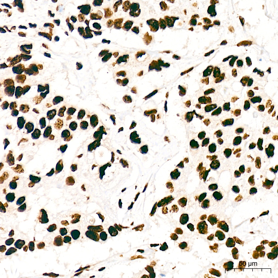
Immunohistochemistry analysis of paraffin-embedded Human breast cancer tissue using SFPQ Rabbit mAb (CAB3494) at a dilution of 1:200 (40x lens). High pressure antigen retrieval was performed with 0.01 M citrate buffer (pH 6.0) prior to IHC staining.
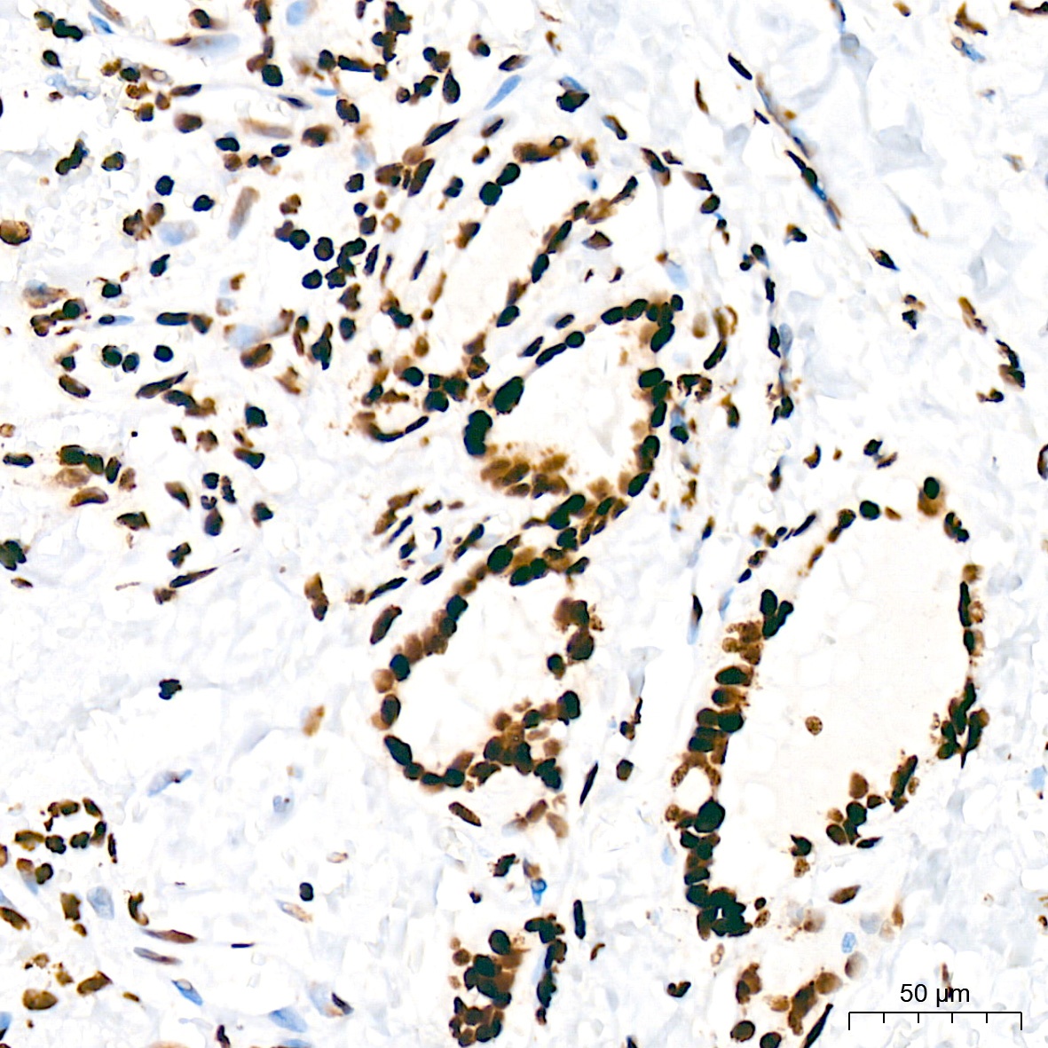
Immunohistochemistry analysis of paraffin-embedded Human thyroid tissue using SFPQ Rabbit mAb (CAB3494) at a dilution of 1:200 (40x lens). High pressure antigen retrieval was performed with 0.01 M citrate buffer (pH 6.0) prior to IHC staining.
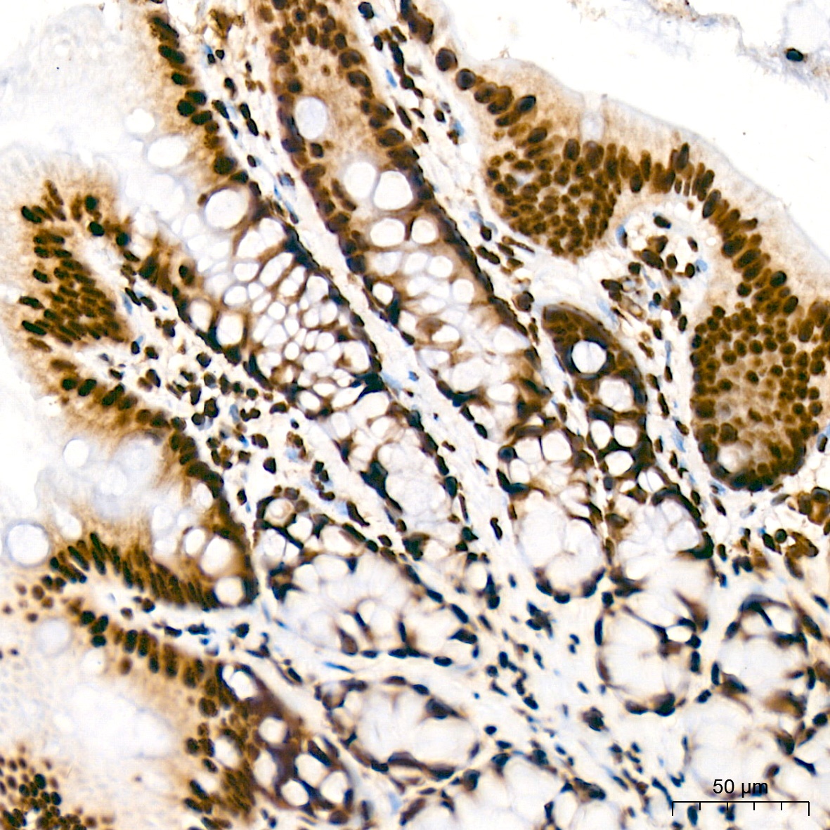
Immunohistochemistry analysis of paraffin-embedded Rat colon tissue using SFPQ Rabbit mAb (CAB3494) at a dilution of 1:200 (40x lens). High pressure antigen retrieval was performed with 0.01 M citrate buffer (pH 6.0) prior to IHC staining.
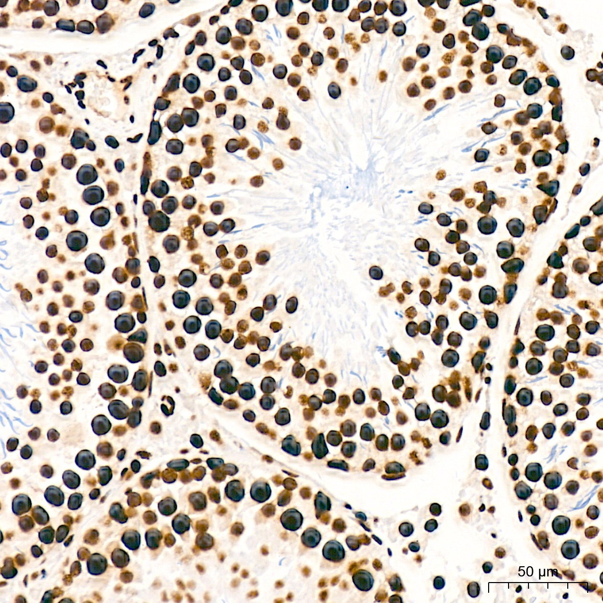
Immunohistochemistry analysis of paraffin-embedded Rat testis tissue using SFPQ Rabbit mAb (CAB3494) at a dilution of 1:200 (40x lens). High pressure antigen retrieval was performed with 0.01 M citrate buffer (pH 6.0) prior to IHC staining.
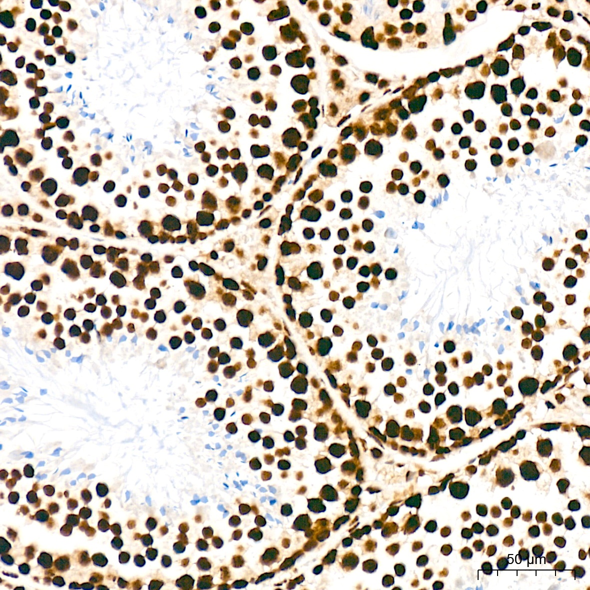
Immunohistochemistry analysis of paraffin-embedded Mouse testis tissue using SFPQ Rabbit mAb (CAB3494) at a dilution of 1:200 (40x lens). High pressure antigen retrieval was performed with 0.01 M citrate buffer (pH 6.0) prior to IHC staining.
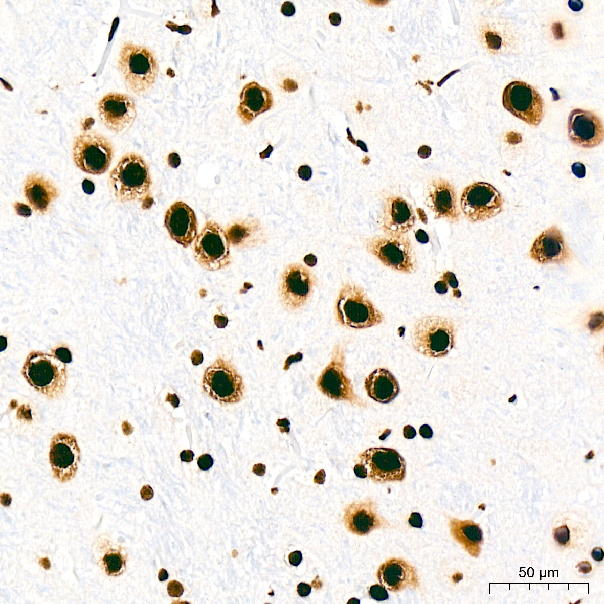
Immunohistochemistry analysis of paraffin-embedded Rat brain tissue using SFPQ Rabbit mAb (CAB3494) at a dilution of 1:200 (40x lens). High pressure antigen retrieval was performed with 0.01 M citrate buffer (pH 6.0) prior to IHC staining.
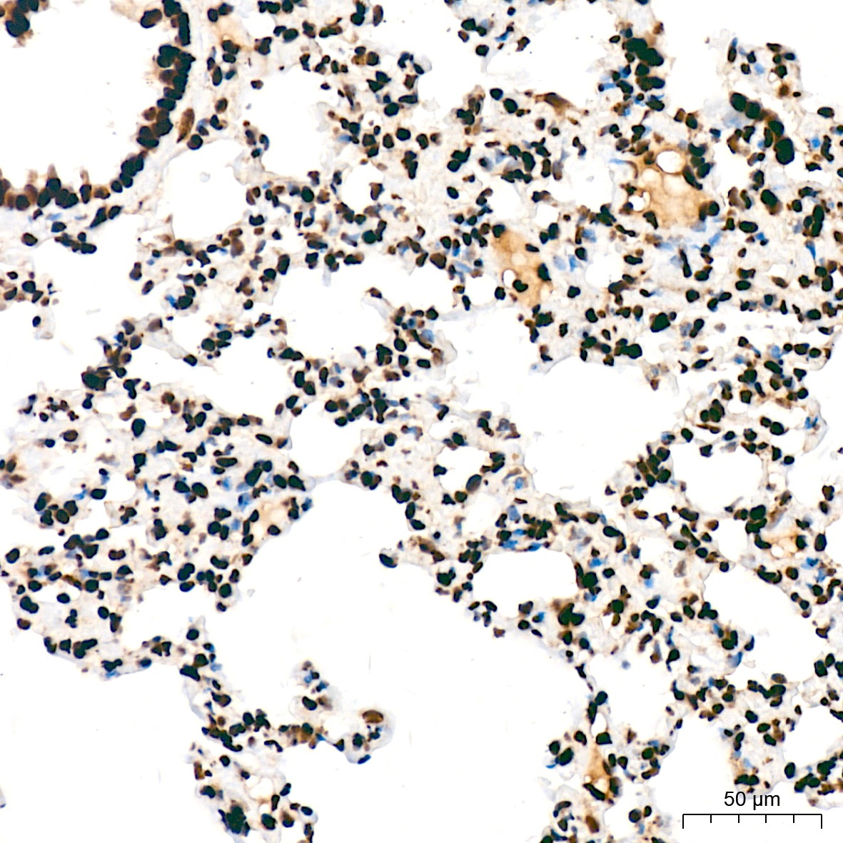
Immunohistochemistry analysis of paraffin-embedded Rat lung tissue using SFPQ Rabbit mAb (CAB3494) at a dilution of 1:200 (40x lens). High pressure antigen retrieval was performed with 0.01 M citrate buffer (pH 6.0) prior to IHC staining.
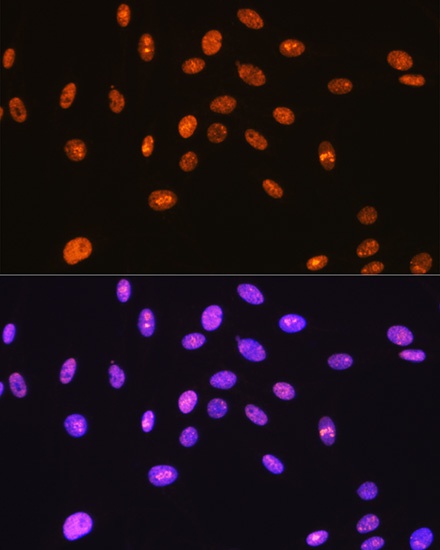
Immunofluorescence analysis of C6 cells using SFPQ Rabbit mAb (CAB3494) at dilution of 1:100 (40x lens). Secondary antibody: Cy3-conjugated Goat anti-Rabbit IgG (H+L) (CABS007) at 1:500 dilution. Blue: DAPI for nuclear staining.
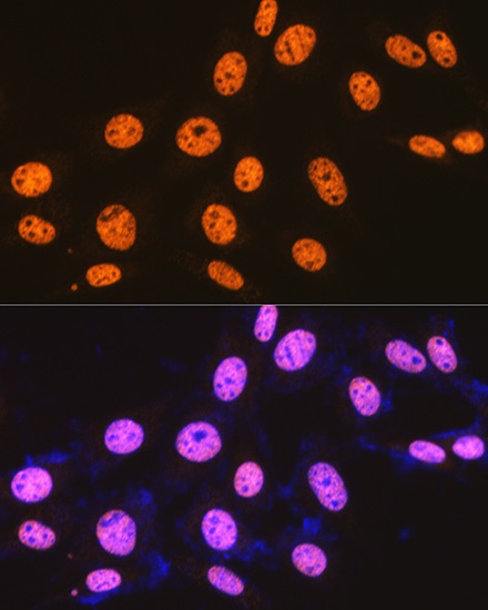
Immunofluorescence analysis of NIH-3T3 cells using SFPQ Rabbit mAb (CAB3494) at dilution of 1:100 (40x lens). Secondary antibody: Cy3-conjugated Goat anti-Rabbit IgG (H+L) (CABS007) at 1:500 dilution. Blue: DAPI for nuclear staining.