The Smad3 Antibody (CAB16913) is a high-quality antibody developed for reliable detection and analysis of target proteins. This antibody, generated in rabbits, exhibits high specificity and sensitivity towards human samples, making it suitable for use in Western blot applications.Smad3 is a crucial mediator of TGF-beta signaling, regulating the expression of genes involved in cell proliferation, differentiation, and apoptosis. Dysregulation of the Smad3 pathway has been implicated in various diseases, including cancer, fibrosis, and immune disorders.
This antibody is validated for use in WB, IF/ICC, ELISA applications and has demonstrated reactivity against Human, Mouse, Rat samples.
Product Name:
Smad3 Antibody
SKU:
CAB16913
Size:
20μL, 100μL
Reactivity:
Human, Mouse, Rat
Immunogen:
Recombinant protein (or fragment).This information is considered to be commercially sensitive.
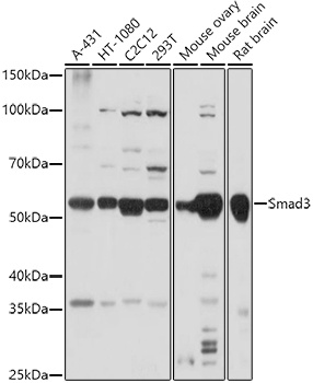
A-431, HT-1080, C2C12, 293T, Mouse ovary, Mouse brain, Rat brain
Cellular Localization:
Cytoplasm, Nucleus.
Calculated MW:
48kDa
Observed MW:
52kDa
The SMAD family of proteins are a group of intracellular signal transducer proteins similar to the gene products of the Drosophila gene 'mothers against decapentaplegic' (Mad) and the C. elegans gene Sma. The SMAD3 protein functions in the transforming growth factor-beta signaling pathway, and transmits signals from the cell surface to the nucleus, regulating gene activity and cell proliferation. This protein forms a complex with other SMAD proteins and binds DNA, functioning both as a transcription factor and tumor suppressor. Mutations in this gene are associated with aneurysms-osteoarthritis syndrome and Loeys-Dietz Syndrome 3.
Purification Method
Affinity purification
Gene ID
4088
RRID
AB_2772307
Buffer Information
Store at -20℃. Avoid freeze / thaw cycles. Buffer: PBS containing 50% glycerol, preserved with proclin300 or sodium azide, pH 7.3.
Western blot analysis of various lysates using (CAB16913) at 1:1000 dilution. Secondary antibody: HRP-conjugated Goat anti-Rabbit IgG (H+L) (CABS014) at 1:10000 dilution. Lysates/proteins: 25μg per lane. Blocking buffer: 3% nonfat dry milk in TBST. Detection: ECL Basic Kit (AbGn00020). Exposure time: 30s.
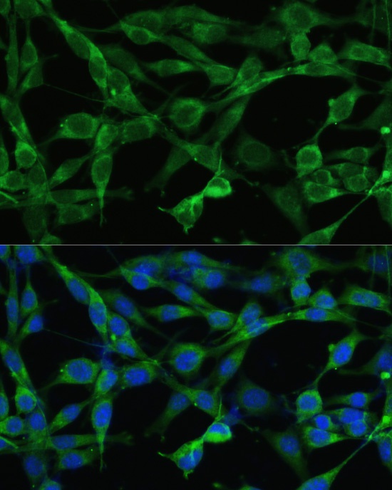
Immunofluorescence analysis of NIH/3T3 cells using Smad3 Rabbit pAb (CAB16913) at dilution of 1:100. Blue: DAPI for nuclear staining.


")