The [KO Validated] SND1 Antibody (CAB5874) is a high-quality antibody developed for reliable detection and analysis of target proteins. This antibody, produced in rabbits, displays high reactivity with human samples and has been validated for use in Western blot applications. By binding specifically to the SND1 protein, researchers can accurately detect and analyze SND1 expression in a variety of cell types, making it an essential tool for studies in molecular biology and virology.
This antibody is validated for use in WB, IHC-P, IF/ICC, IP, ELISA applications and has demonstrated reactivity against Human, Mouse, Rat samples.
Product Name:
[KO Validated] SND1 Antibody
SKU:
CAB5874
Size:
20μL, 100μL
Reactivity:
Human, Mouse, Rat
Conjugate:
Unconjugated
Immunogen:
Recombinant protein (or fragment).This information is considered to be commercially sensitive.
Tested Applications:
WBIHC-PIF/ICCIPELISA
Recommended Dilution:
WB
1:500 - 1:2000
IHC-P
1:50 - 1:200
IF/ICC
1:50 - 1:200
IP
0.5μg-4μg antibody for 200μg-400μg extracts of whole cells
ELISA
Recommended starting concentration is 1 μg/mL. Please optimize the concentration based on your specific assay requirements.
Synonyms:
p100, TDRD11, Tudor-SN, D1
Positive Sample:
HeLa, Jurkat, HL-60, MCF7, 293T, Mouse liver, Rat liver
Cellular Localization:
Cytoplasm, Melanosome, Nucleus.
Calculated MW:
102kDa
Observed MW:
120kDa
This gene encodes a transcriptional co-activator that interacts with the acidic domain of Epstein-Barr virus nuclear antigen 2 (EBNA 2), a transcriptional activator that is required for B-lymphocyte transformation. Other transcription factors that interact with this protein are signal transducers and activators of transcription, STATs. This protein is also thought to be essential for normal cell growth. A similar protein in mammals and other organisms is a component of the RNA-induced silencing complex (RISC).
Purification Method
Affinity purification
Gene ID
27044
RRID
AB_2766623
Buffer Information
Store at -20℃. Avoid freeze / thaw cycles. Buffer: PBS containing 50% glycerol, preserved with proclin300 or sodium azide, pH 7.3.
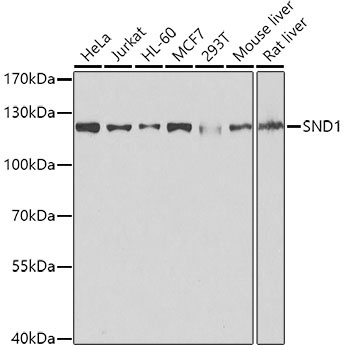
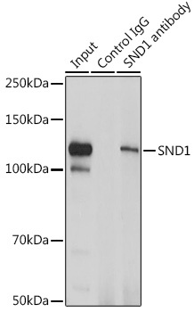
Western blot analysis of various lysates using [KO Validated] SND1 Rabbit pAb (CAB5874) at 1:1000 dilution. Secondary antibody: HRP-conjugated Goat anti-Rabbit IgG (H+L) (CABS014) at 1:10000 dilution. Lysates/proteins: 25μg per lane. Blocking buffer: 3% nonfat dry milk in TBST. Detection: ECL Basic Kit (AbGn00020). Exposure time: 1s.
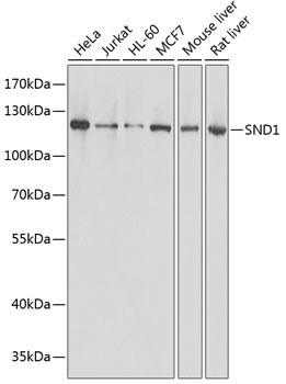
Western blot analysis of various lysates using [KO Validated] SND1 Rabbit pAb (CAB5874) at 1:1000 dilution. Secondary antibody: HRP-conjugated Goat anti-Rabbit IgG (H+L) (CABS014) at 1:10000 dilution. Lysates/proteins: 25μg per lane. Blocking buffer: 3% nonfat dry milk in TBST. Detection: ECL Basic Kit (AbGn00020). Exposure time: 1s.
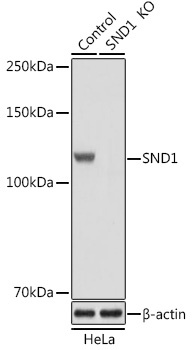
Western blot analysis of lysates from wild type (WT) and SND1 knockout (KO) HeLa cells, using [KO Validated] SND1 Rabbit pAb (CAB5874) at 1:1000 dilution. Secondary antibody: HRP-conjugated Goat anti-Rabbit IgG (H+L) (CABS014) at 1:10000 dilution. Lysates/proteins: 25μg per lane. Blocking buffer: 3% nonfat dry milk in TBST. Detection: ECL Basic Kit (AbGn00020). Exposure time: 1s.
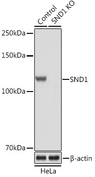
Western blot analysis of lysates from wild type (WT) and SND1 knockout (KO) HeLa cells, using [KO Validated] SND1 Rabbit pAb (CAB5874) at 1:3000 dilution. Secondary antibody: HRP-conjugated Goat anti-Rabbit IgG (H+L) (CABS014) at 1:10000 dilution. Lysates/proteins: 25μg per lane. Blocking buffer: 3% nonfat dry milk in TBST. Detection: ECL Basic Kit (AbGn00020). Exposure time: 1s.
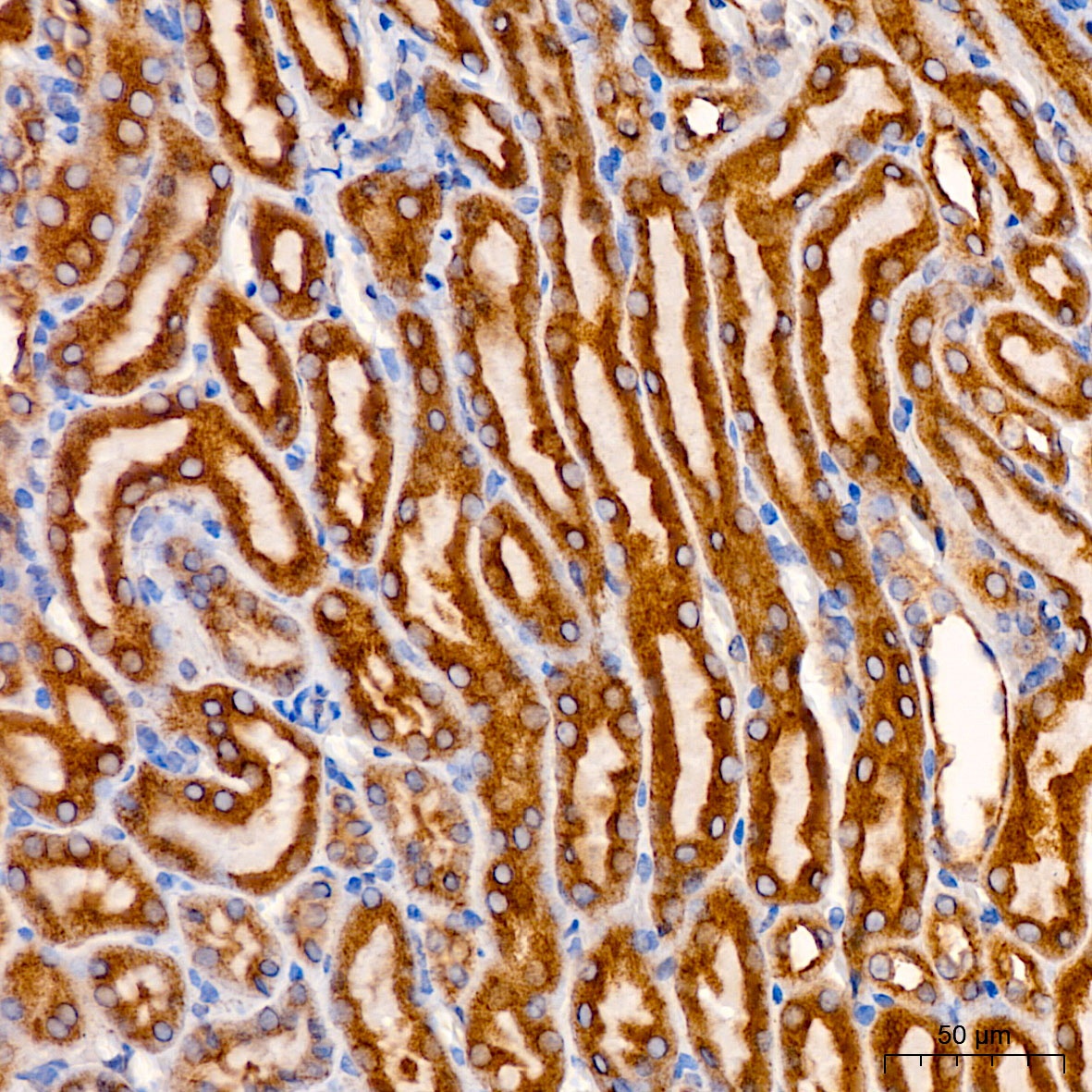
Immunohistochemistry analysis of paraffin-embedded Mouse kidney using [KO Validated] SND1 Rabbit pAb (CAB5874) at dilution of 1:200 (40x lens). High pressure antigen retrieval performed with 0.01M Citrate buffer (pH 6.0) prior to IHC staining.
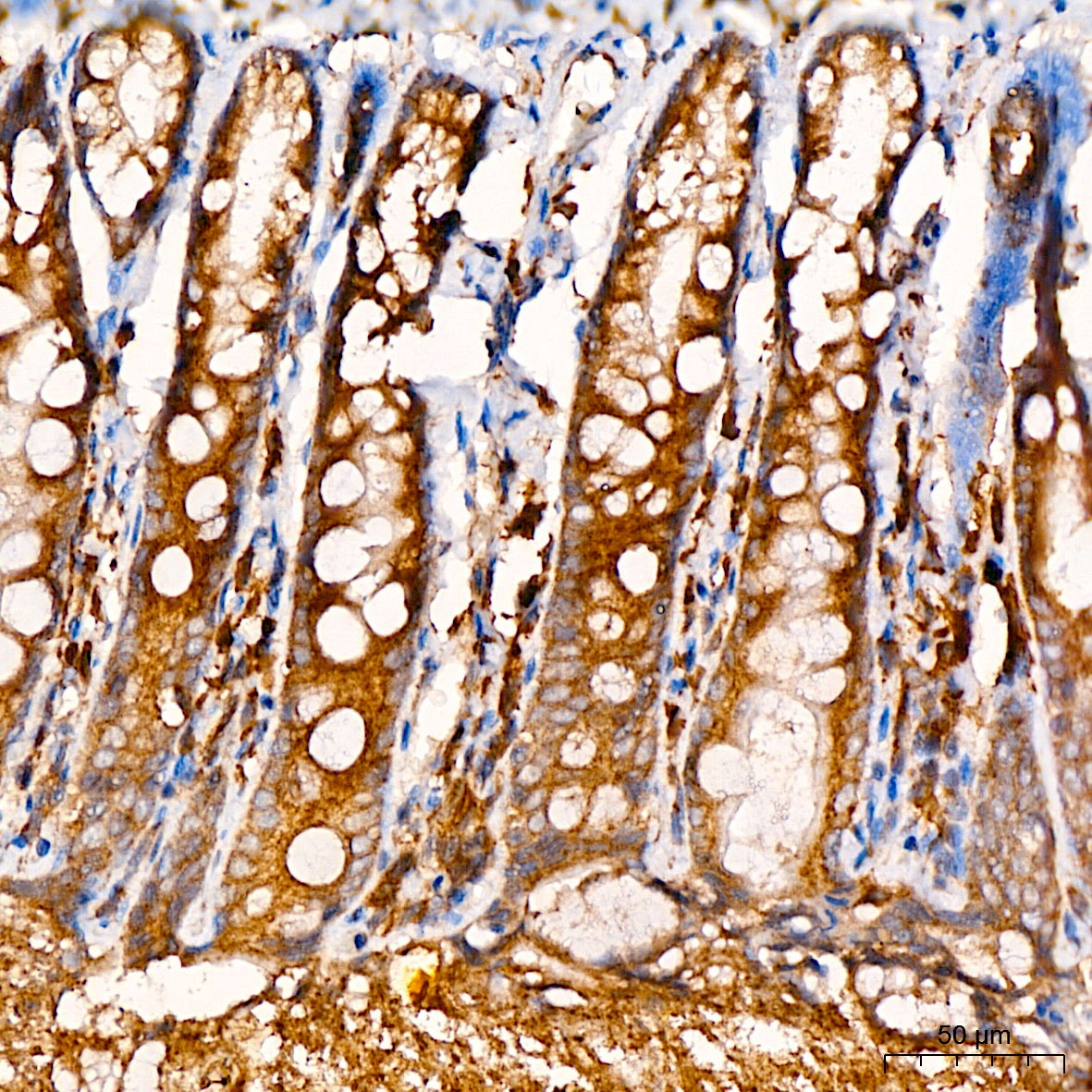
Immunohistochemistry analysis of paraffin-embedded Rat colon using [KO Validated] SND1 Rabbit pAb (CAB5874) at dilution of 1:200 (40x lens). High pressure antigen retrieval performed with 0.01M Citrate buffer (pH 6.0) prior to IHC staining.
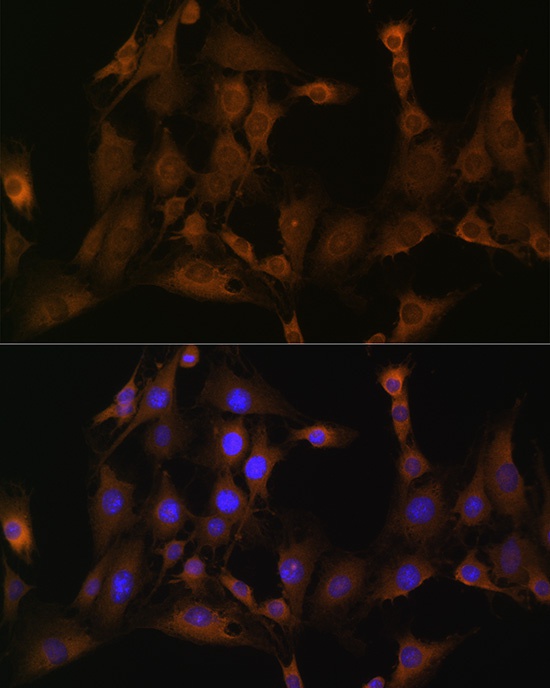
Immunofluorescence analysis of C6 cells using SND1 Rabbit pAb (CAB5874) at dilution of 1:100 (40x lens). Secondary antibody: Cy3-conjugated Goat anti-Rabbit IgG (H+L) (CABS007) at 1:500 dilution. Blue: DAPI for nuclear staining.
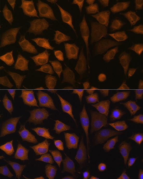
Immunofluorescence analysis of L929 cells using SND1 Rabbit pAb (CAB5874) at dilution of 1:100 (40x lens). Secondary antibody: Cy3-conjugated Goat anti-Rabbit IgG (H+L) (CABS007) at 1:500 dilution. Blue: DAPI for nuclear staining.
Immunoprecipitation analysis of 200 μg extracts of Jurkat cells using 3 μg SND1 antibody (CAB5874). Western blot was performed from the immunoprecipitate using SND1 antibody (CAB5874) at a dilution of 1:1000.