The SOX9 Monoclonal Antibody (CAB19710) is a high-quality antibody developed for reliable detection and analysis of target proteins. This antibody, generated in rabbits, is highly specific to human samples and is validated for use in a variety of applications, including Western blotting.SOX9 is known for its involvement in various biological processes, including chondrogenesis, sex determination, and stem cell maintenance. Dysregulation of SOX9 has been implicated in numerous diseases, such as skeletal disorders, campomelic dysplasia, and certain types of cancer.
This antibody is validated for use in WB, IHC-P, IF/ICC, IP, ELISA, IF-P applications and has demonstrated reactivity against Human, Mouse, Rat samples.
Product Name:
SOX9 Monoclonal Antibody
SKU:
CAB19710
Size:
20μL, 100μL
Reactivity:
Human, Mouse, Rat
Clone Number:
ARC0190
Conjugate:
Unconjugated
Immunogen:
Recombinant protein (or fragment).This information is considered to be commercially sensitive.
0.5μg-4μg antibody for 200μg-400μg extracts of whole cells
IF/ICC
1:200 - 1:2000
IF-P
1:200 - 1:2000
IHC-P
1:1000 - 1:4000
ELISA
Recommended starting concentration is 1 μg/mL. Please optimize the concentration based on your specific assay requirements.
Synonyms:
CMD1, SRA1, CMPD1, SRXX2, SRXY10, SOX9
Positive Sample:
SW480, HCT 116
Cellular Localization:
Nucleus.
Calculated MW:
56kDa
Observed MW:
75kDa
The protein encoded by this gene recognizes the sequence CCTTGAG along with other members of the HMG-box class DNA-binding proteins. It acts during chondrocyte differentiation and, with steroidogenic factor 1, regulates transcription of the anti-Muellerian hormone (AMH) gene. Deficiencies lead to the skeletal malformation syndrome campomelic dysplasia, frequently with sex reversal.
Purification Method
Affinity purification
Gene ID
6662
RRID
AB_2862748
Buffer Information
Store at -20℃. Avoid freeze / thaw cycles. Buffer: PBS containing 50% glycerol and 0.05% BSA, preserved with proclin300 or sodium azide, pH 7.3.
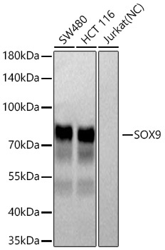
Western blot analysis of various lysates using SOX9 Rabbit mAb (CAB19710) at 1:1000 dilution incubated at room temperature for 1.5 hours. Secondary antibody: HRP-conjugated Goat anti-Rabbit IgG (H+L) (CABS014) at 1:10000 dilution. Lysates/proteins: 25 μg per lane. Blocking buffer: 3% nonfat dry milk in TBST. Detection: ECL Basic Kit (AbGn00020). Negative control (NC): Jurkat Exposure time: 5s.
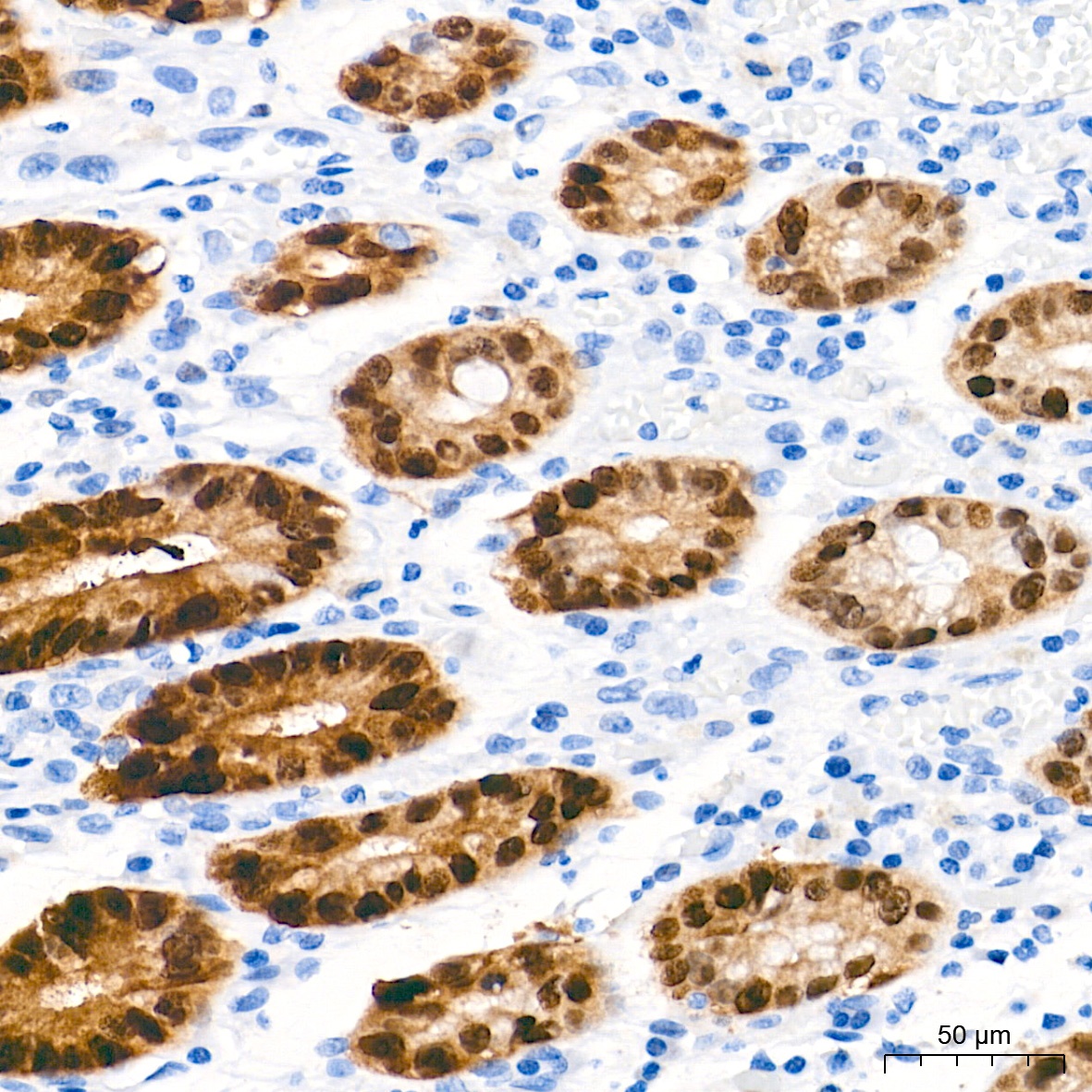
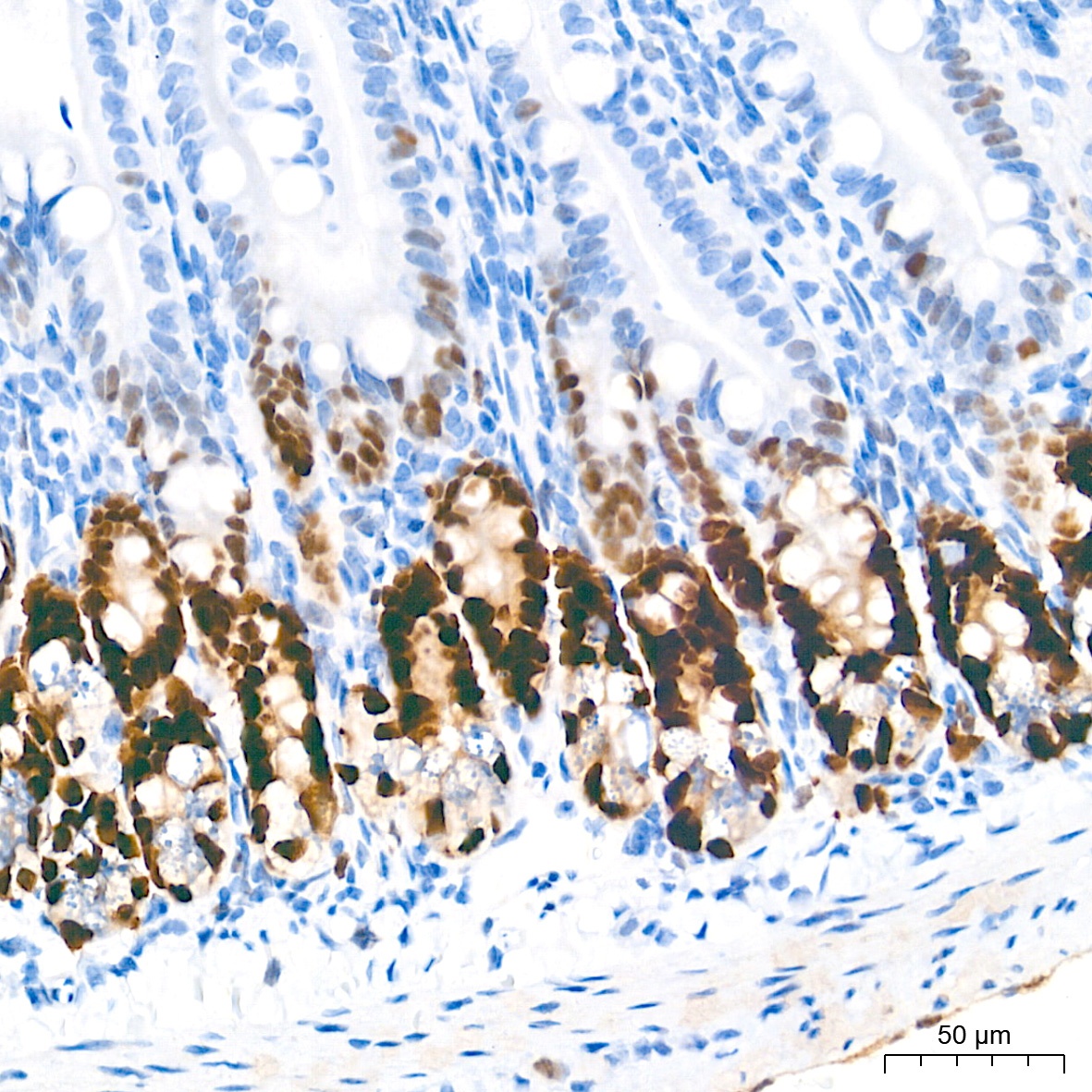
Immunohistochemistry analysis of paraffin-embedded Human colon tissue using SOX9 Rabbit mAb (CAB19710) at a dilution of 1:1000 (40x lens). High pressure antigen retrieval performed with 0.01M Tris-EDTA Buffer (pH 9.0) prior to IHC staining.
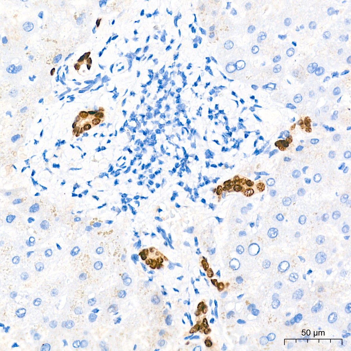
Immunohistochemistry analysis of paraffin-embedded Human liver tissue using SOX9 Rabbit mAb (CAB19710) at a dilution of 1:1000 (40x lens). High pressure antigen retrieval performed with 0.01M Tris-EDTA Buffer (pH 9.0) prior to IHC staining.
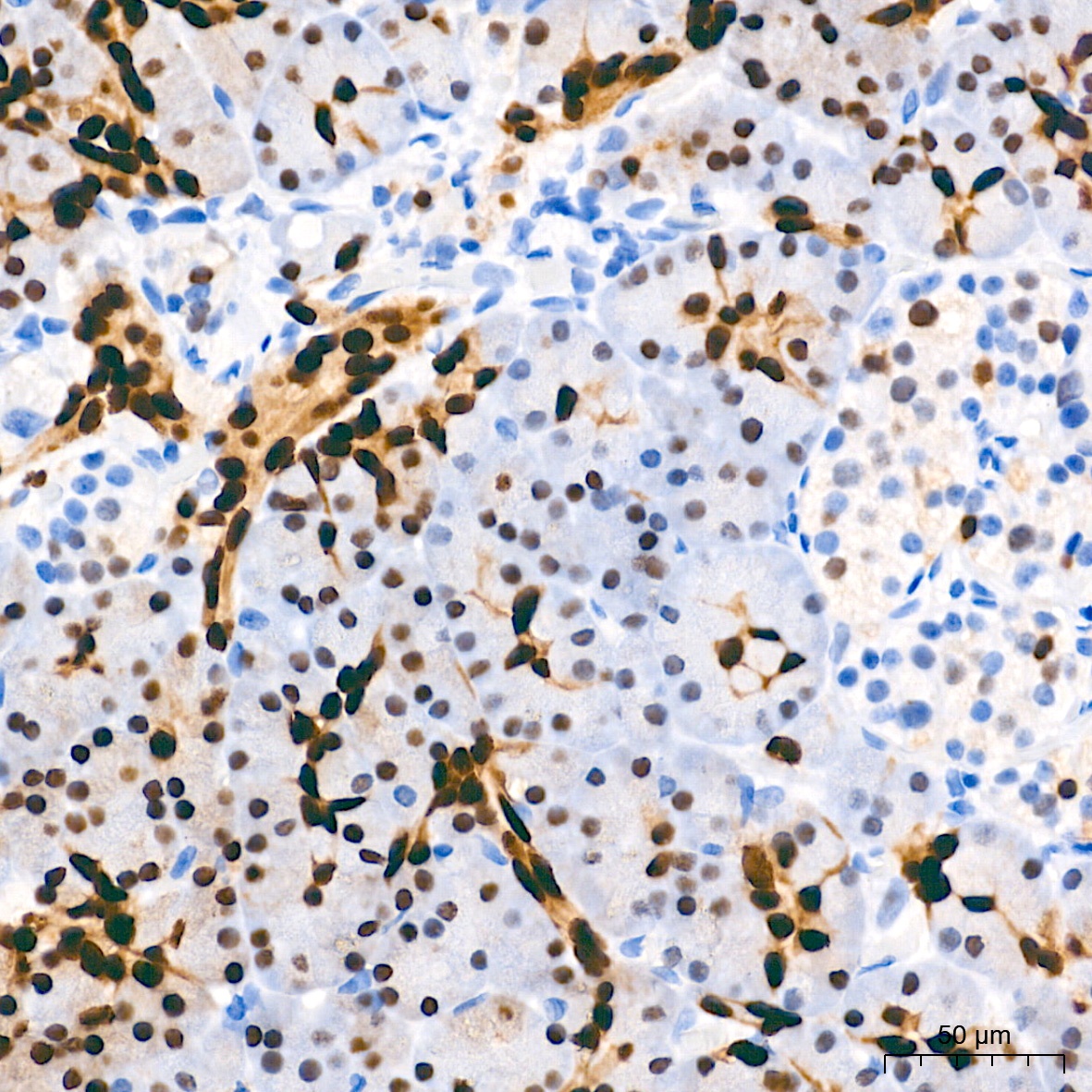
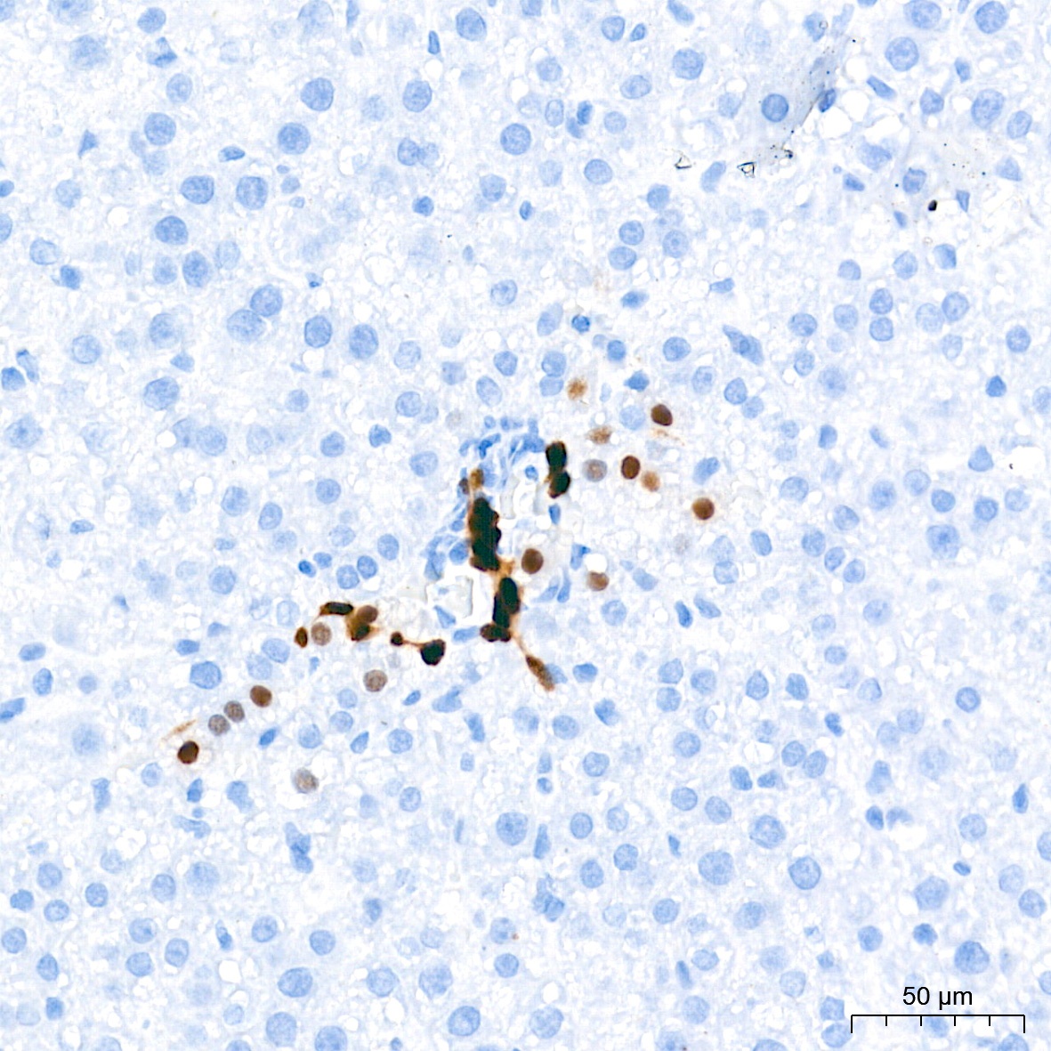
Immunohistochemistry analysis of paraffin-embedded Human pancreas tissue using SOX9 Rabbit mAb (CAB19710) at a dilution of 1:1000 (40x lens). High pressure antigen retrieval performed with 0.01M Tris-EDTA Buffer (pH 9.0) prior to IHC staining.
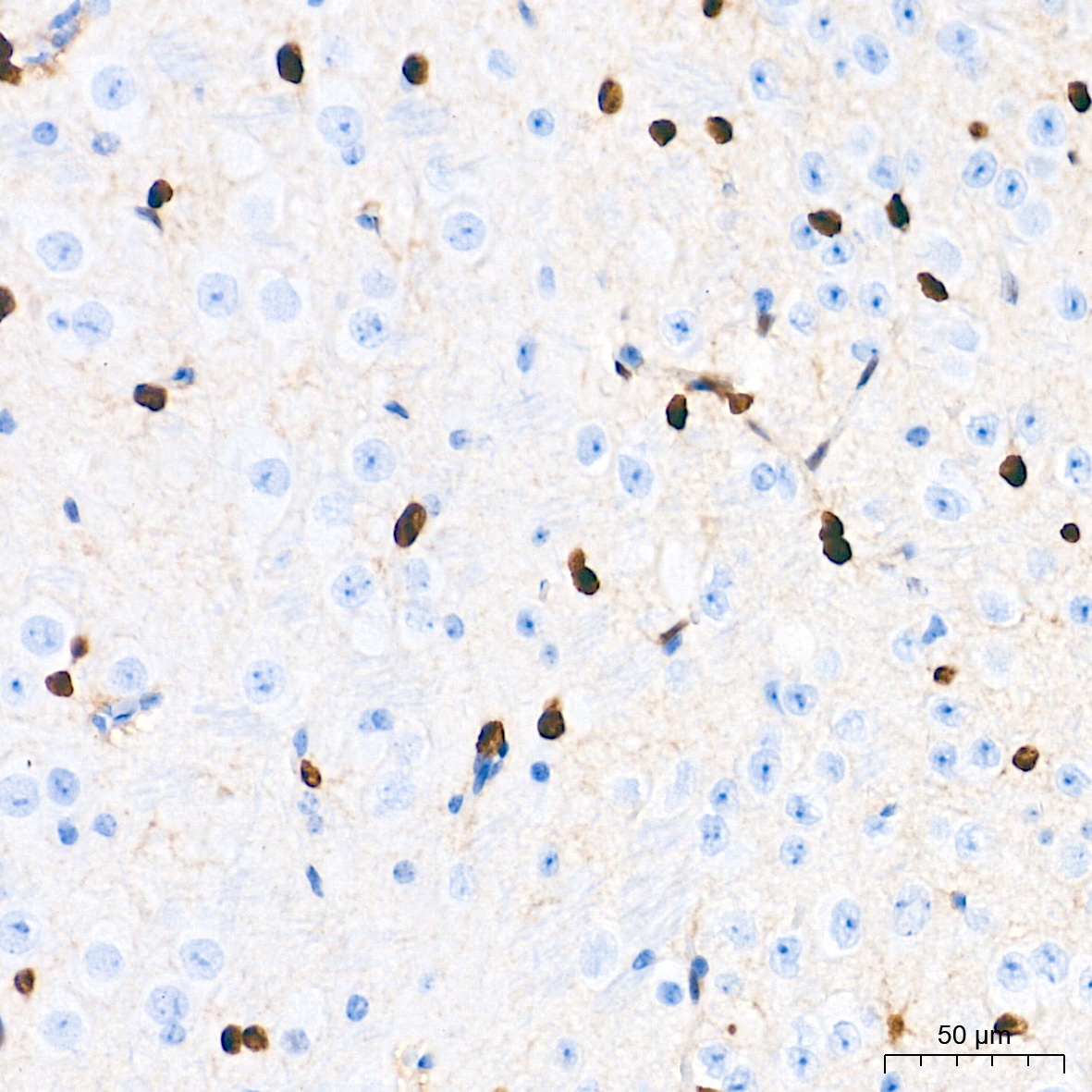
Immunohistochemistry analysis of paraffin-embedded Mouse brain tissue using SOX9 Rabbit mAb (CAB19710) at a dilution of 1:1000 (40x lens). High pressure antigen retrieval performed with 0.01M Tris-EDTA Buffer (pH 9.0) prior to IHC staining.
Immunohistochemistry analysis of paraffin-embedded Mouse intestin tissue using SOX9 Rabbit mAb (CAB19710) at a dilution of 1:1000 (40x lens). High pressure antigen retrieval performed with 0.01M Tris-EDTA Buffer (pH 9.0) prior to IHC staining.
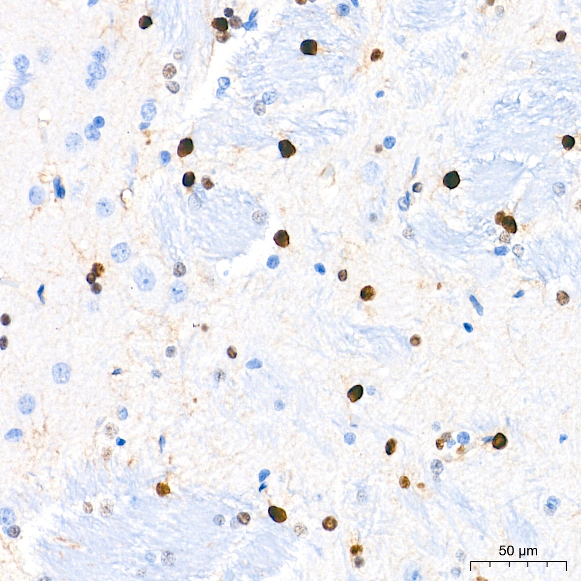
Immunohistochemistry analysis of paraffin-embedded Rat brain tissue using SOX9 Rabbit mAb (CAB19710) at a dilution of 1:1000 (40x lens). High pressure antigen retrieval performed with 0.01M Tris-EDTA Buffer (pH 9.0) prior to IHC staining.
Immunohistochemistry analysis of paraffin-embedded Rat liver tissue using SOX9 Rabbit mAb (CAB19710) at a dilution of 1:1000 (40x lens). High pressure antigen retrieval performed with 0.01M Tris-EDTA Buffer (pH 9.0) prior to IHC staining.
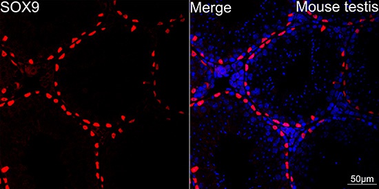
Confocal imaging of paraffin-embedded Mouse testis using SOX9 Rabbit mAb (CAB19710, dilution 1:200) followed by a further incubation with Cy3 Goat Anti-Rabbit IgG (H+L) (CABS007, dilution 1:500) (Red). DAPI was used for nuclear staining (Blue). Objective: 40x.Perform high pressure antigen retrieval with 0.01M citrate buffer (pH 6.0) prior to IF staining.
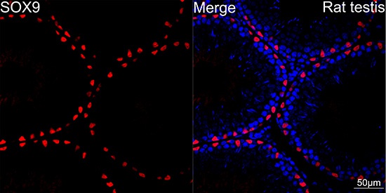
Confocal imaging of paraffin-embedded Rat testis using SOX9 Rabbit mAb (CAB19710, dilution 1:200) followed by a further incubation with Cy3 Goat Anti-Rabbit IgG (H+L) (CABS007, dilution 1:500) (Red). DAPI was used for nuclear staining (Blue). Objective: 40x.Perform high pressure antigen retrieval with 0.01M citrate buffer (pH 6.0) prior to IF staining.
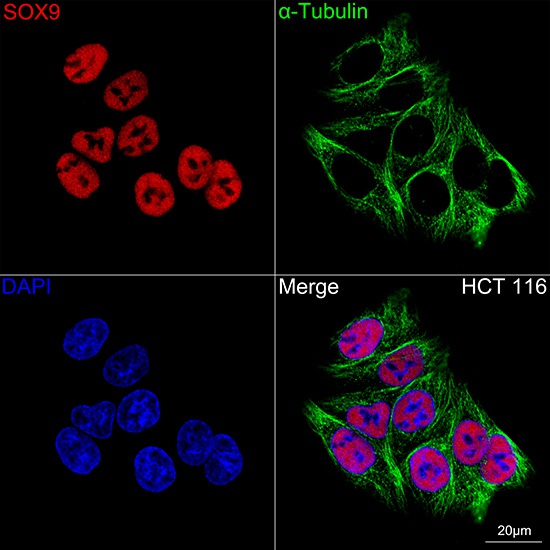
Confocal imaging of HCT 116 cells using SOX9 Rabbit mAb (CAB19710, dilution 1:200) followed by a further incubation with Cy3 Goat Anti-Rabbit IgG (H+L) (CABS007, dilution 1:500) (Red). The cells were counterstained with α-Tubulin Mouse mAb (AC012, dilution 1:400) followed by incubation with ABflo® 488-conjugated Goat Anti-Mouse IgG (H+L) Ab (CABS076, dilution 1:500) (Green). DAPI was used for nuclear staining (Blue). Objective: 100x.