The SOX9 Monoclonal Antibody (CAB19710) is a high-quality antibody developed for reliable detection and analysis of target proteins. The protein encoded by this gene recognizes the sequence CCTTGAG along with other members of the HMG-box class DNA-binding proteins. It acts during chondrocyte differentiation and, with steroidogenic factor 1, regulates transcription of the anti-Muellerian hormone (AMH) gene. Deficiencies lead to the skeletal malformation syndrome campomelic dysplasia, frequently with sex reversal. RRID AB_2862748 Gene ID 6662 Swiss Prot Synonym CMD1; SRA1; CMPD1; SRXX2; SRXY10; SOX9
This antibody is validated for use in WB, IHC-P, IF/ICC, IP, ELISA, IF-P applications and has demonstrated reactivity against Human, Mouse, Rat samples.
Product Name:
SOX9 Monoclonal Antibody
SKU:
CAB19710
Size:
100μL, 20μL
Reactivity:
Human, Mouse, Rat
Clone Number:
ARC0190
Conjugate:
Unconjugated
Immunogen:
Recombinant protein (or fragment).This information is considered to be commercially sensitive.
Tested Applications:
WBIHC-PIF/ICCIPELISAIF-P
Recommended Dilution:
WB
1:1000 - 1:2000
IP
0.5μg-4μg antibody for 200μg-400μg extracts of whole cells
IF
/
ICC
1:200 - 1:2000
IF-P
1:200 - 1:2000
IHC-P
1:1000 - 1:4000
ELISA
Recommended starting concentration is 1 μg/mL. Please optimize the concentration based on your specific assay requirements.
Synonyms:
CMD1, SRA1, CMPD1, SRXX2, SRXY10, SOX9
Positive Sample:
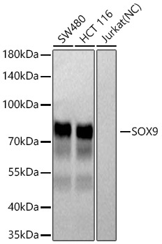
SW480, HCT 116
Cellular Localization:
Nucleus.
Calculated MW:
56kDa
Observed MW:
75kDa
The protein encoded by this gene recognizes the sequence CCTTGAG along with other members of the HMG-box class DNA-binding proteins. It acts during chondrocyte differentiation and, with steroidogenic factor 1, regulates transcription of the anti-Muellerian hormone (AMH) gene. Deficiencies lead to the skeletal malformation syndrome campomelic dysplasia, frequently with sex reversal. RRID AB_2862748 Gene ID 6662 Swiss Prot Synonym CMD1; SRA1; CMPD1; SRXX2; SRXY10; SOX9
Purification Method:
Affinity purification
Gene ID:
6662
RRID:
AB_2862748
Buffer Information:
Store at -20℃. Avoid freeze / thaw cycles. Buffer: PBS containing 50% glycerol and 0.05% BSA, preserved with proclin300 or sodium azide, pH 7.3.
Western blot analysis of various lysates using SOX9 Rabbit mAb (CAB19710) at 1:1000 dilution incubated at room temperature for 1.5 hours. Secondary antibody: HRP-conjugated Goat anti-Rabbit IgG (H+L) (AS014) at 1:10000 dilution. Lysates/proteins: 25 μg per lane. Blocking buffer: 3% nonfat dry milk in TBST. Detection: ECL Basic Kit (AbGn00020). Negative control (NC): Jurkat Exposure time: 5s.
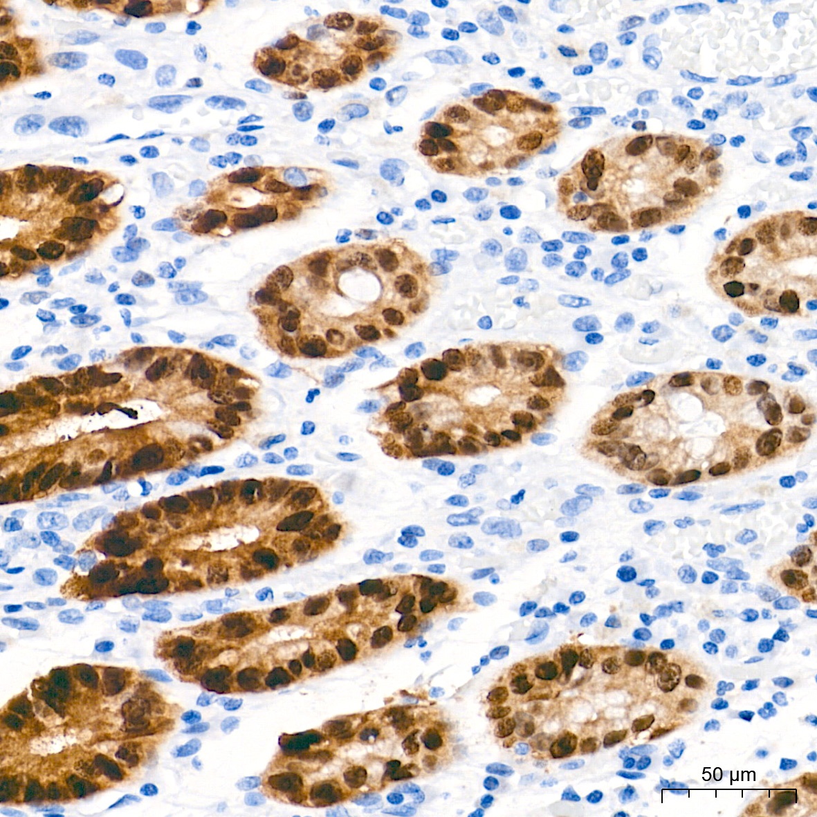
Immunohistochemistry analysis of paraffin-embedded Human colon tissue using SOX9 Rabbit mAb (CAB19710) at a dilution of 1:1000 (40x lens). High pressure antigen retrieval performed with 0.01M Tris-EDTA Buffer (pH 9.0) prior to IHC staining.
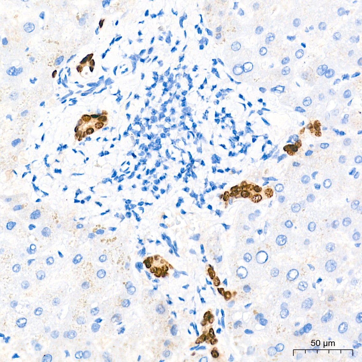
Immunohistochemistry analysis of paraffin-embedded Human liver tissue using SOX9 Rabbit mAb (CAB19710) at a dilution of 1:1000 (40x lens). High pressure antigen retrieval performed with 0.01M Tris-EDTA Buffer (pH 9.0) prior to IHC staining.
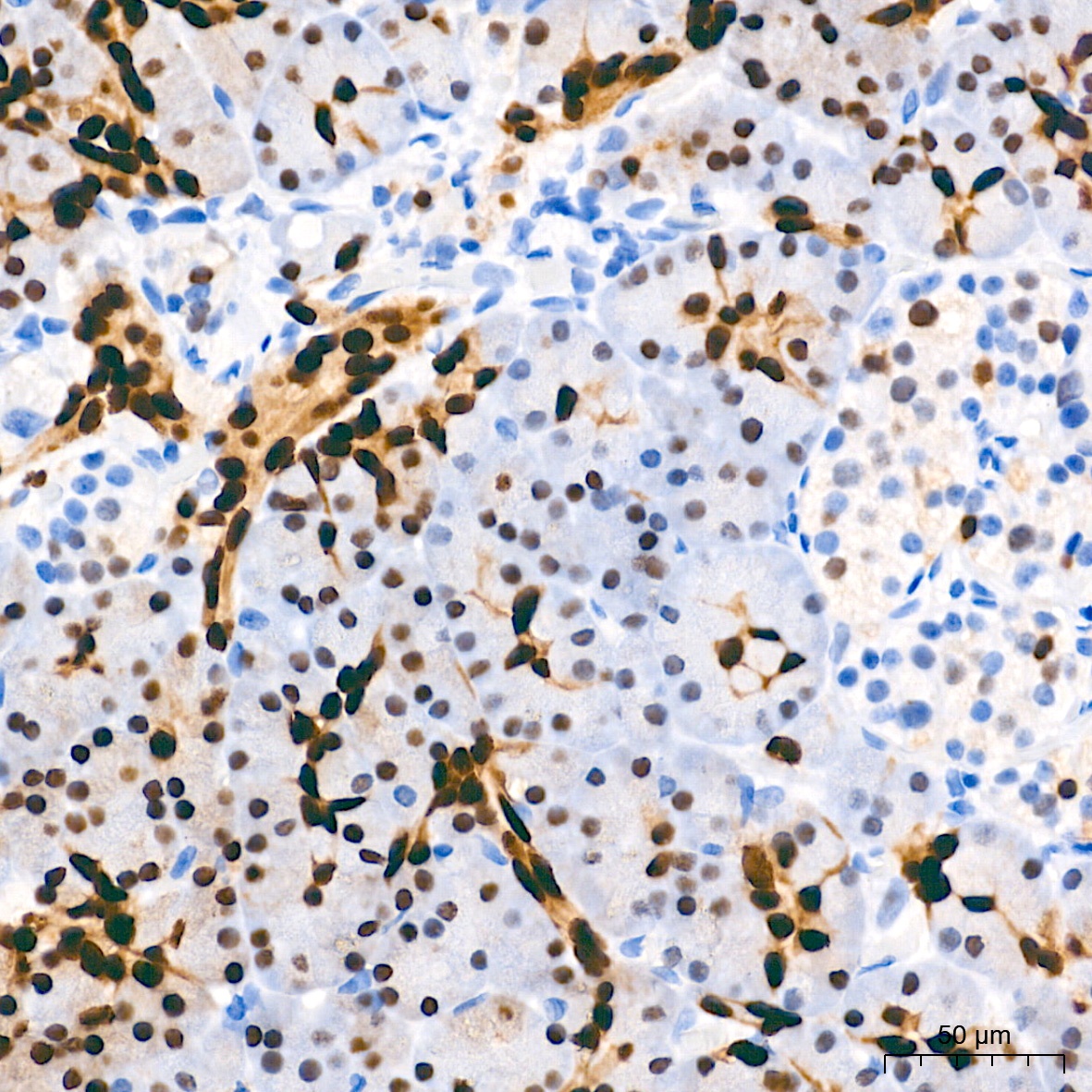
Immunohistochemistry analysis of paraffin-embedded Human pancreas tissue using SOX9 Rabbit mAb (CAB19710) at a dilution of 1:1000 (40x lens). High pressure antigen retrieval performed with 0.01M Tris-EDTA Buffer (pH 9.0) prior to IHC staining.
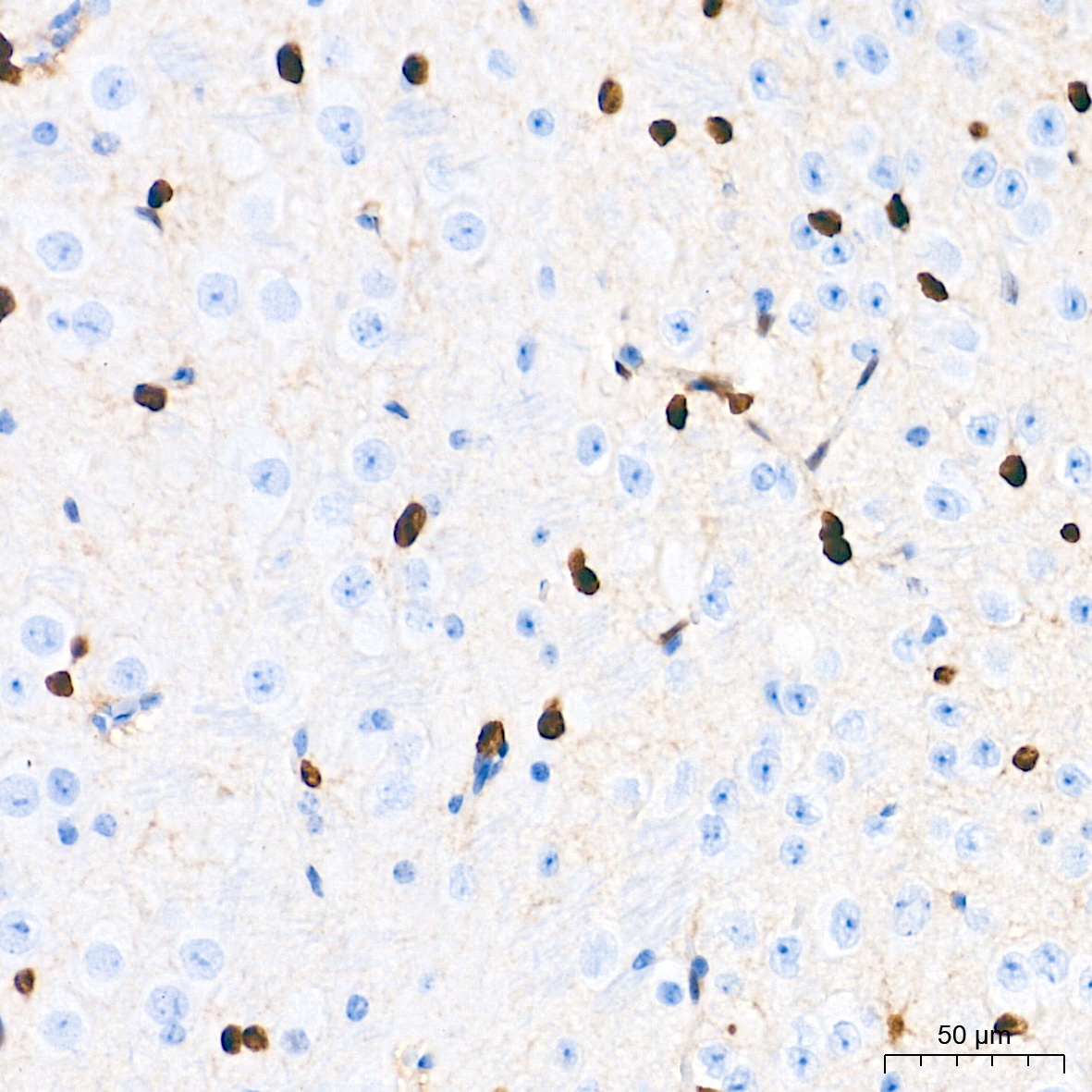
Immunohistochemistry analysis of paraffin-embedded Mouse brain tissue using SOX9 Rabbit mAb (CAB19710) at a dilution of 1:1000 (40x lens). High pressure antigen retrieval performed with 0.01M Tris-EDTA Buffer (pH 9.0) prior to IHC staining.
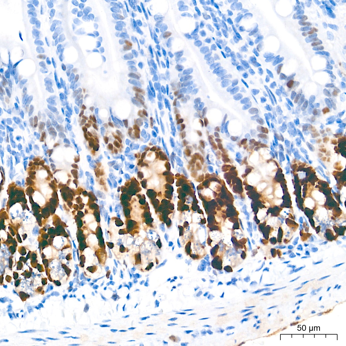
Immunohistochemistry analysis of paraffin-embedded Mouse intestin tissue using SOX9 Rabbit mAb (CAB19710) at a dilution of 1:1000 (40x lens). High pressure antigen retrieval performed with 0.01M Tris-EDTA Buffer (pH 9.0) prior to IHC staining.
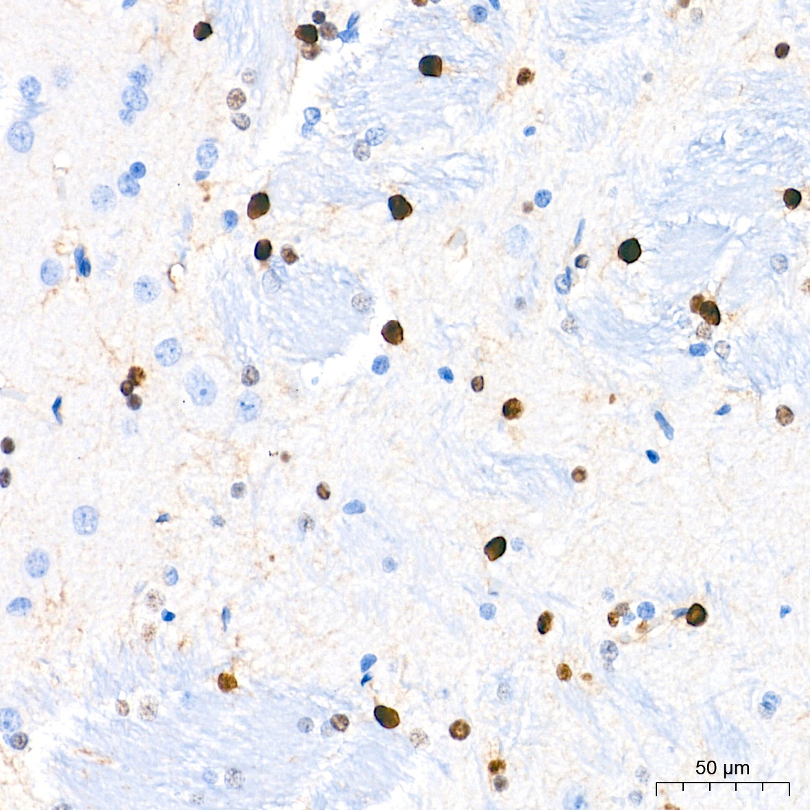
Immunohistochemistry analysis of paraffin-embedded Rat brain tissue using SOX9 Rabbit mAb (CAB19710) at a dilution of 1:1000 (40x lens). High pressure antigen retrieval performed with 0.01M Tris-EDTA Buffer (pH 9.0) prior to IHC staining.
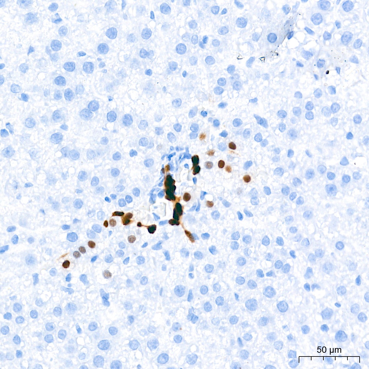
Immunohistochemistry analysis of paraffin-embedded Rat liver tissue using SOX9 Rabbit mAb (CAB19710) at a dilution of 1:1000 (40x lens). High pressure antigen retrieval performed with 0.01M Tris-EDTA Buffer (pH 9.0) prior to IHC staining.
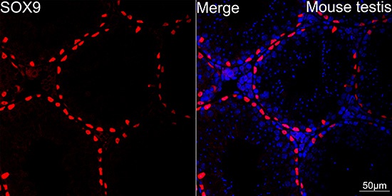
Confocal imaging of paraffin-embedded Mouse testis using SOX9 Rabbit mAb (CAB19710, dilution 1:200) followed by a further incubation with Cy3 Goat Anti-Rabbit IgG (H+L) (AS007, dilution 1:500) (Red). DAPI was used for nuclear staining (Blue). Objective: 40x.Perform high pressure antigen retrieval with 0.01M citrate buffer (pH 6.0) prior to IF staining.
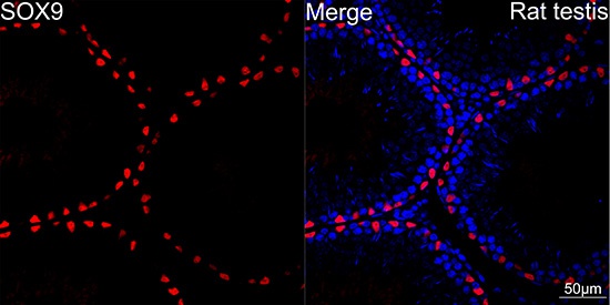
Confocal imaging of paraffin-embedded Rat testis using SOX9 Rabbit mAb (CAB19710, dilution 1:200) followed by a further incubation with Cy3 Goat Anti-Rabbit IgG (H+L) (AS007, dilution 1:500) (Red). DAPI was used for nuclear staining (Blue). Objective: 40x.Perform high pressure antigen retrieval with 0.01M citrate buffer (pH 6.0) prior to IF staining.
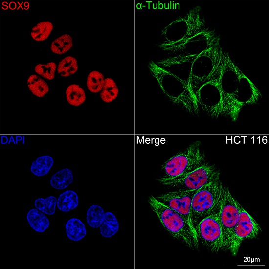
Confocal imaging of HCT 116 cells using SOX9 Rabbit mAb (CAB19710, dilution 1:200) followed by a further incubation with Cy3 Goat Anti-Rabbit IgG (H+L) (AS007, dilution 1:500) (Red). The cells were counterstained with α-Tubulin Mouse mAb (AC012, dilution 1:400) followed by incubation with ABflo® 488-conjugated Goat Anti-Mouse IgG (H+L) Ab (AS076, dilution 1:500) (Green). DAPI was used for nuclear staining (Blue). Objective: 100x.