The STIM2 Antibody (CAB17743) is a high-quality antibody developed for reliable detection and analysis of target proteins. This antibody, generated in rabbits, exhibits high reactivity with human samples and is validated for use in Western blot applications. By specifically binding to the STIM2 protein, this antibody enables precise detection and analysis in various cell types, making it ideal for investigations in immunology, neuroscience, and cancer research.
This antibody is validated for use in WB, IHC-P, IF/ICC, ELISA applications and has demonstrated reactivity against Human, Mouse, Rat samples.
Product Name:
STIM2 Antibody
SKU:
CAB17743
Size:
20μL, 100μL
Reactivity:
Human, Mouse, Rat
Conjugate:
Unconjugated
Immunogen:
Recombinant protein (or fragment).This information is considered to be commercially sensitive.
Recommended starting concentration is 1 μg/mL. Please optimize the concentration based on your specific assay requirements.
Synonyms:
STIM2
Positive Sample:
293T
Cellular Localization:
Endoplasmic Reticulum Membrane, Single-Pass Type I Membrane Protein.
Calculated MW:
84kDa
Observed MW:
110kDa
This gene is a member of the stromal interaction molecule (STIM) family and likely arose, along with related family member STIM1, from a common ancestral gene. The encoded protein functions to regulate calcium concentrations in the cytosol and endoplasmic reticulum, and is involved in the activation of plasma membrane Orai Ca(2+) entry channels. This gene initiates translation from a non-AUG (UUG) start site. A signal peptide is cleaved from the resulting protein. Multiple transcript variants result from alternative splicing.
Purification Method
Affinity purification
Gene ID
57620
RRID
AB_2772431
Buffer Information
Store at -20℃. Avoid freeze / thaw cycles. Buffer: PBS with 0.01% thimerosal,50% glycerol,pH7.3.
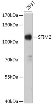
Western blot analysis of lysates from 293T cells, using STIM2 Rabbit pAb (CAB17743) at 1:1000 dilution. Secondary antibody: HRP-conjugated Goat anti-Rabbit IgG (H+L) (CABS014) at 1:10000 dilution. Lysates/proteins: 25μg per lane. Blocking buffer: 3% nonfat dry milk in TBST. Detection: ECL Basic Kit (AbGn00020). Exposure time: 30s.
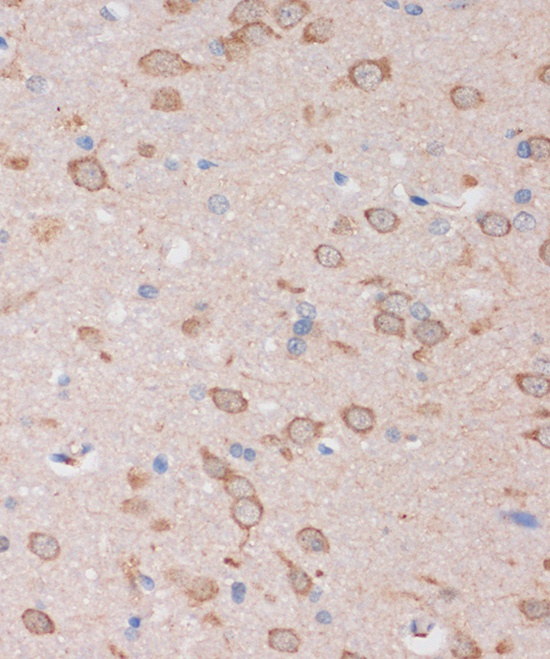
Immunohistochemistry analysis of paraffin-embedded Rat brain using STIM2 Rabbit pAb (CAB17743) at dilution of 1:100 (40x lens). Microwave antigen retrieval performed with 0.01M PBS Buffer (pH 7.2) prior to IHC staining.
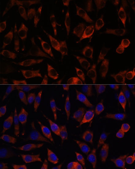
Immunofluorescence analysis of L929 cells using STIM2 Rabbit pAb (CAB17743) at dilution of 1:100. Secondary antibody: Cy3-conjugated Goat anti-Rabbit IgG (H+L) (CABS007) at 1:500 dilution. Blue: DAPI for nuclear staining.
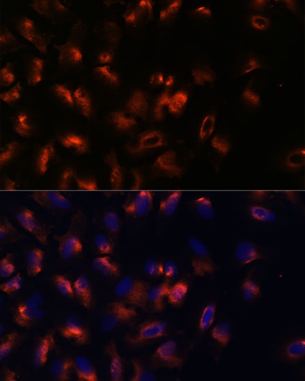
Immunofluorescence analysis of U-2 OS cells using STIM2 Rabbit pAb (CAB17743) at dilution of 1:100. Secondary antibody: Cy3-conjugated Goat anti-Rabbit IgG (H+L) (CABS007) at 1:500 dilution. Blue: DAPI for nuclear staining.