The SUFU Antibody (CAB6757) is a high-quality antibody developed for reliable detection and analysis of target proteins. This antibody, produced in rabbits, shows high reactivity with human samples and is validated for use in Western blot applications. By binding to the SUFU protein, this antibody enables researchers to detect and analyze SUFU expression in various cell types, making it an essential component in studies related to developmental biology, cancer research, and stem cell regulation.SUFU is a critical regulator of the hedgehog signaling pathway, which plays a crucial role in embryonic development, tissue patterning, and stem cell maintenance.
This antibody is validated for use in WB, IHC-P, IF/ICC, ELISA applications and has demonstrated reactivity against Human, Mouse, Rat samples.
Product Name:
SUFU Antibody
SKU:
CAB6757
Size:
20μL, 100μL
Reactivity:
Human, Mouse, Rat
Conjugate:
Unconjugated
Immunogen:
Recombinant protein (or fragment).This information is considered to be commercially sensitive.
Recommended starting concentration is 1 μg/mL. Please optimize the concentration based on your specific assay requirements.
Synonyms:
SUFUH, JBTS32, SUFUXL, PRO1280, SUFU
Positive Sample:
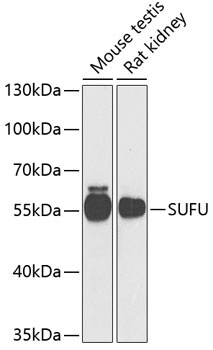
Mouse testis, Rat kidney
Cellular Localization:
Cytoplasm, Nucleus.
Calculated MW:
54kDa
Observed MW:
55kDa
The Hedgehog signaling pathway plays an important role in early human development. The pathway is a signaling cascade that plays a role in pattern formation and cellular proliferation during development. This gene encodes a negative regulator of the hedgehog signaling pathway. Defects in this gene are a cause of medulloblastoma. Alternative splicing results in multiple transcript variants.
Purification Method
Affinity purification
Gene ID
51684
RRID
AB_2767341
Buffer Information
Store at -20℃. Avoid freeze / thaw cycles. Buffer: PBS containing 50% glycerol, preserved with proclin300 or sodium azide, pH 7.3.
Western blot analysis of various lysates using SUFU Rabbit pAb (CAB6757) at 1:1000 dilution. Secondary antibody: HRP-conjugated Goat anti-Rabbit IgG (H+L) (CABS014) at 1:10000 dilution. Lysates/proteins: 25μg per lane. Blocking buffer: 3% nonfat dry milk in TBST. Detection: ECL Basic Kit (AbGn00020). Exposure time: 90s.
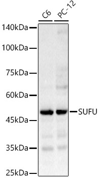
Western blot analysis of various lysates, using SUFU Rabbit pAb (CAB6757) at 1:1000 dilution. Secondary antibody: HRP-conjugated Goat anti-Rabbit IgG (H+L) (CABS014) at 1:10000 dilution. Lysates/proteins: 25μg per lane. Blocking buffer: 3% nonfat dry milk in TBST. Detection: ECL Enhanced Kit (AbGn00021). Exposure time: 10s.
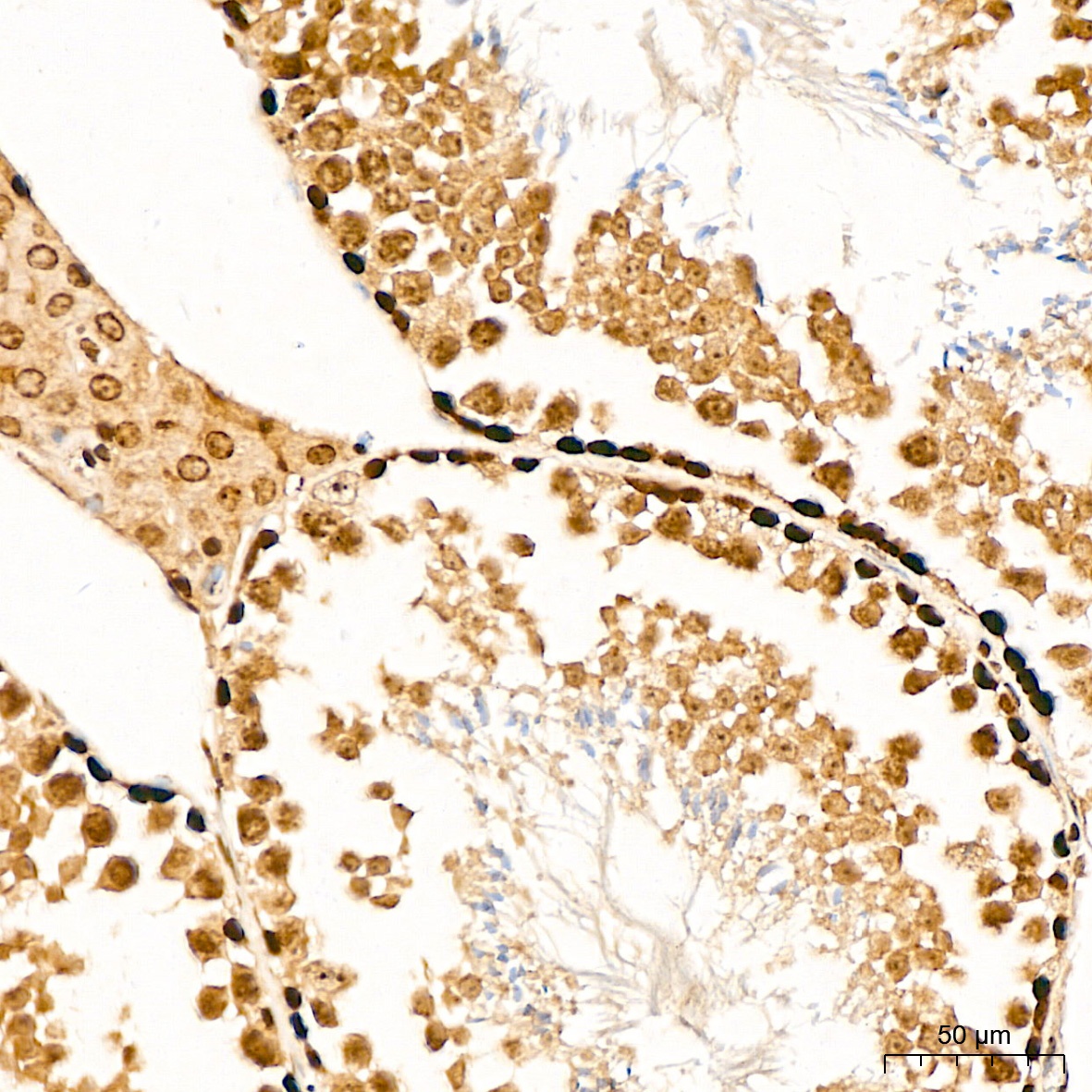
Immunohistochemistry analysis of paraffin-embedded Mouse testis tissue using SUFU Rabbit pAb (CAB6757) at a dilution of 1:400 (40x lens). High pressure antigen retrieval was performed with 0.01 M citrate buffer (pH 6.0) prior to IHC staining.
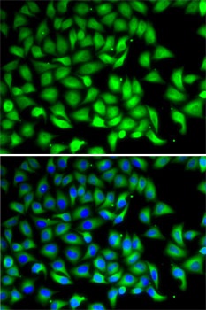
Immunofluorescence analysis of MCF7 cells using SUFU Rabbit pAb (CAB6757). Secondary antibody: Cy3-conjugated Goat anti-Rabbit IgG (H+L) (CABS007) at 1:500 dilution. Blue: DAPI for nuclear staining.