The Synaptophysin Antibody (CAB6344) is a high-quality antibody developed for reliable detection and analysis of target proteins. This antibody, produced in rabbits, demonstrates high reactivity with human samples and is specifically validated for use in Western blot applications.Synaptophysin is a critical protein involved in neurotransmission, playing a key role in synaptic vesicle fusion and neurotransmitter release. Research on synaptophysin is essential for understanding synaptic function, neuronal development, and synaptic plasticity, making this antibody an essential component for studies in neuroscience, neurology, and neurodegenerative diseases.
This antibody is validated for use in WB, IHC-P, IF/ICC, IP, ELISA, IF-P applications and has demonstrated reactivity against Human, Mouse, Rat samples.
Product Name:
Synaptophysin Antibody
SKU:
CAB6344
Size:
20μL, 100μL
Reactivity:
Human, Mouse, Rat
Conjugate:
Unconjugated
Immunogen:
Recombinant protein (or fragment).This information is considered to be commercially sensitive.
This gene encodes an integral membrane protein of small synaptic vesicles in brain and endocrine cells. The protein also binds cholesterol and is thought to direct targeting of vesicle-associated membrane protein 2 (synaptobrevin) to intracellular compartments. Mutations in this gene are associated with an X-linked form of cognitive disability.
Purification Method
Affinity purification
Gene ID
6855
RRID
AB_2766946
Buffer Information
Store at -20℃. Avoid freeze / thaw cycles. Buffer: PBS with 0.09% Sodium azide,50% glycerol,pH7.3.
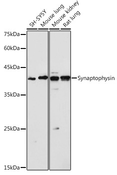
Western blot analysis of various lysates using Synaptophysin Rabbit pAb (CAB6344) at 1:1000 dilution. Secondary antibody: HRP-conjugated Goat anti-Rabbit IgG (H+L) (CABS014) at 1:10000 dilution. Lysates/proteins: 25μg per lane. Blocking buffer: 3% nonfat dry milk in TBST. Detection: ECL Basic Kit (AbGn00020). Exposure time: 1s.
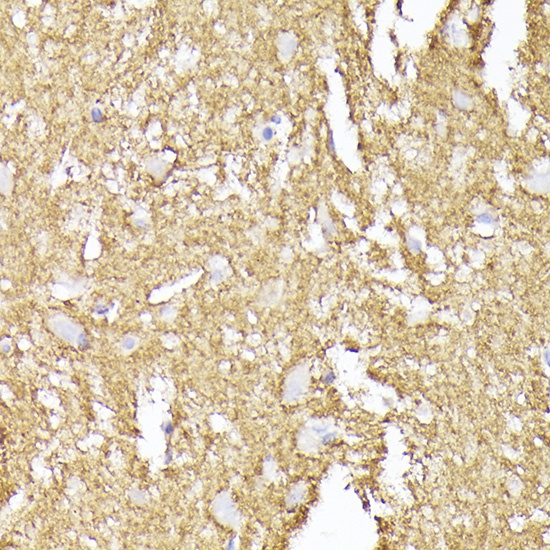
Immunohistochemistry analysis of paraffin-embedded Human brain using Synaptophysin Rabbit pAb (CAB6344) at dilution of 1:200 (40x lens). High pressure antigen retrieval performed with 0.01M Citrate buffer (pH 6.0) prior to IHC staining.
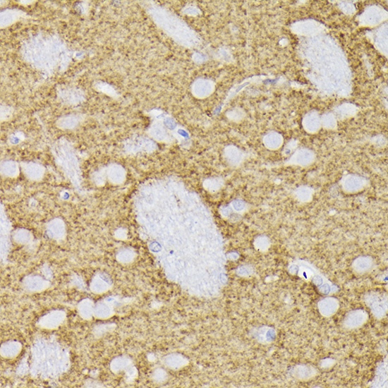
Immunohistochemistry analysis of paraffin-embedded Rat brain using Synaptophysin Rabbit pAb (CAB6344) at dilution of 1:200 (40x lens). High pressure antigen retrieval performed with 0.01M Citrate buffer (pH 6.0) prior to IHC staining.
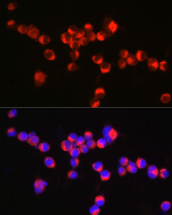
Immunofluorescence analysis of Neuro-2a cells using Synaptophysin Rabbit pAb (CAB6344) at dilution of 1:100 (40x lens). Secondary antibody: Cy3-conjugated Goat anti-Rabbit IgG (H+L) (CABS007) at 1:500 dilution. Blue: DAPI for nuclear staining.
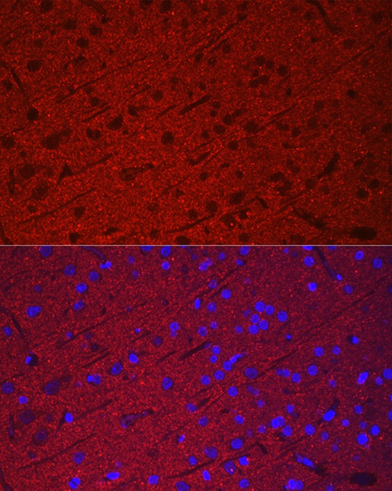
Immunofluorescence analysis of paraffin-embedded rat brain using Synaptophysin Rabbit pAb (CAB6344) at dilution of 1:100 (40x lens). Secondary antibody: Cy3-conjugated Goat anti-Rabbit IgG (H+L) (CABS007) at 1:500 dilution. Blue: DAPI for nuclear staining.
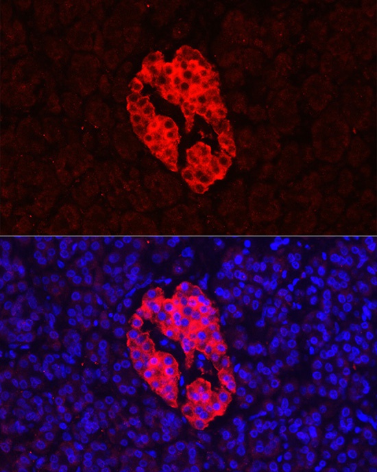
Immunofluorescence analysis of paraffin-embedded human pancreas using Synaptophysin Rabbit pAb (CAB6344) at dilution of 1:100 (40x lens). Secondary antibody: Cy3-conjugated Goat anti-Rabbit IgG (H+L) (CABS007) at 1:500 dilution. Blue: DAPI for nuclear staining.
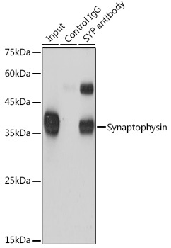
Immunoprecipitation analysis of 600 μg extracts of Mouse brain cells using 3 μg Synaptophysin antibody (CAB6344). Western blot was performed from the immunoprecipitate using Synaptophysin antibody (CAB6344) at a dilution of 1:1000.