The TAF15 Antibody (CAB8465) is a high-quality antibody developed for reliable detection and analysis of target proteins. This antibody, generated in rabbits, demonstrates high reactivity with human samples and has been extensively validated for use in Western blotting applications. By specifically binding to the TAF15 protein, researchers can effectively detect and analyze TAF15 expression in a variety of cell types, making it an essential component for studies in molecular biology and cancer research.TAF15 is a multifunctional protein that plays a crucial role in gene expression regulation and has been implicated in the development and progression of various diseases, including cancer.
This antibody is validated for use in WB, IHC-P, IF/ICC, IP, ELISA applications and has demonstrated reactivity against Human samples.
Product Name:
TAF15 Antibody
SKU:
CAB8465
Size:
20μL, 100μL
Reactivity:
Human
Conjugate:
Unconjugated
Immunogen:
Recombinant protein (or fragment).This information is considered to be commercially sensitive.
0.5μg-4μg antibody for 200μg-400μg extracts of whole cells
ELISA
Recommended starting concentration is 1 μg/mL. Please optimize the concentration based on your specific assay requirements.
Synonyms:
Npl3, RBP56, TAF2N, TAFII68, TAF15
Positive Sample:
SKOV3, Jurkat
Cellular Localization:
Cytoplasm, Nucleus.
Calculated MW:
62kDa
Observed MW:
80kDa
This gene encodes a member of the TET family of RNA-binding proteins. The encoded protein plays a role in RNA polymerase II gene transcription as a component of a distinct subset of multi-subunit transcription initiation factor TFIID complexes. Translocations involving this gene play a role in acute leukemia and extraskeletal myxoid chondrosarcoma, and mutations in this gene may play a role in amyotrophic lateral sclerosis. Alternatively spliced transcript variants encoding multiple isoforms have been observed for this gene.
Purification Method
Affinity purification
Gene ID
8148
RRID
AB_2772511
Buffer Information
Store at -20℃. Avoid freeze / thaw cycles. Buffer: PBS containing 50% glycerol, preserved with proclin300 or sodium azide, pH 7.3.
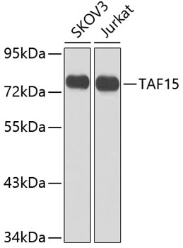
Western blot analysis of various lysates using TAF15 Rabbit pAb (CAB8465) at 1:1000 dilution. Secondary antibody: HRP-conjugated Goat anti-Rabbit IgG (H+L) (CABS014) at 1:10000 dilution. Lysates/proteins: 25μg per lane. Blocking buffer: 3% nonfat dry milk in TBST. Detection: ECL Enhanced Kit (AbGn00021). Exposure time: 5s.
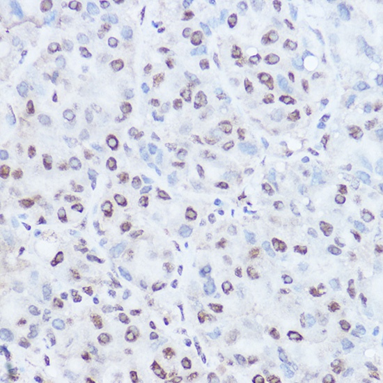
Immunohistochemistry analysis of paraffin-embedded Human liver cancer using TAF15 Rabbit pAb (CAB8465) at dilution of 1:100 (40x lens). High pressure antigen retrieval performed with 0.01M Citrate buffer (pH 6.0) prior to IHC staining.