The TBCCD1 Antibody (CAB18143) is a high-quality antibody developed for reliable detection and analysis of target proteins. The antibody is raised in rabbits and is highly reactive with human samples, making it ideal for use in various research applications such as Western blotting.TBCCD1 is known to be involved in processes related to cell division and chromosome segregation, making it an important target for studying cellular biology and potential therapeutic interventions. The antibody binds specifically to TBCCD1 protein, enabling researchers to detect and analyze its expression in different cell types.
This antibody is validated for use in WB, IHC-P, ELISA applications and has demonstrated reactivity against Human, Mouse, Rat samples.
Product Name:
TBCCD1 Antibody
SKU:
CAB18143
Size:
20μL, 100μL
Reactivity:
Human, Mouse, Rat
Immunogen:
Synthetic peptide. This information is considered to be commercially sensitive.
Recommended starting concentration is 1 μg/mL. Please optimize the concentration based on your specific assay requirements.
Synonyms:
TBCCD1
Positive Sample:
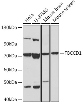
HeLa, U-87MG, Mouse brain, Mouse spleen
Cellular Localization:
Cytoplasm, Spindle Pole Centrosome.
Calculated MW:
64kDa
Observed MW:
70kDa
Involved in several processes, including maintenance of Golgi location; maintenance of centrosome location; and regulation of cell shape. Located in spindle pole centrosome.
Purification Method
Affinity purification
Gene ID
55171
RRID
AB_2861934
Buffer Information
Store at -20℃. Avoid freeze / thaw cycles. Buffer: PBS with 0.01% thimerosal,50% glycerol,pH7.3.
Western blot analysis of various lysates using TBCCD1 Rabbit pAb (CAB18143) at 1:1000 dilution. Secondary antibody: HRP-conjugated Goat anti-Rabbit IgG (H+L) (CABS014) at 1:10000 dilution. Lysates/proteins: 25μg per lane. Blocking buffer: 3% nonfat dry milk in TBST. Detection: ECL Basic Kit (AbGn00020). Exposure time: 60s.
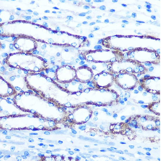
Immunohistochemistry analysis of paraffin-embedded Mouse kidney using TBCCD1 Rabbit pAb (CAB18143) at dilution of 1:100 (40x lens). Microwave antigen retrieval performed with 0.01M PBS Buffer (pH 7.2) prior to IHC staining.
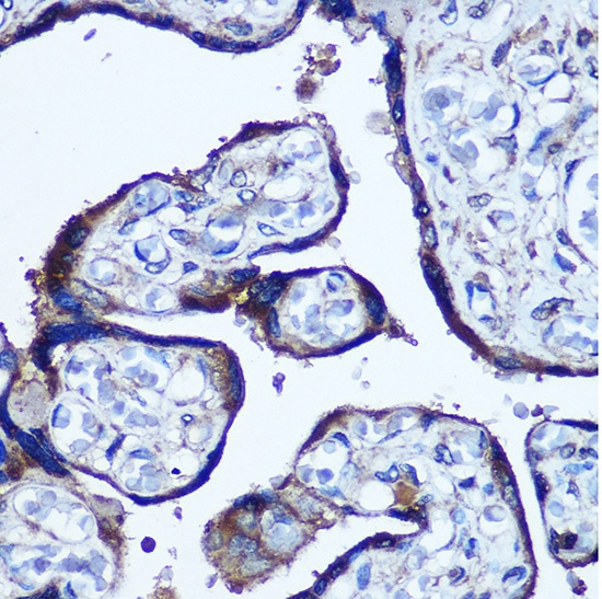
Immunohistochemistry analysis of paraffin-embedded Human placenta using TBCCD1 Rabbit pAb (CAB18143) at dilution of 1:100 (40x lens). Microwave antigen retrieval performed with 0.01M PBS Buffer (pH 7.2) prior to IHC staining.