The TEAD1 Monoclonal Antibody (CAB5092) is a high-quality antibody developed for reliable detection and analysis of target proteins. This antibody, generated in rabbits, exhibits high reactivity with human samples and has been validated for use in applications such as Western blot and immunohistochemistry.The TEAD family of transcription factors are key regulators of gene expression and play a crucial role in various cellular processes, including cell growth, differentiation, and development. By targeting TEAD1, TEAD2, TEAD3, and TEAD4, researchers can gain insights into their functions in different cell types and biological contexts, making this antibody an essential tool for studies in molecular biology and cancer research.
This antibody is validated for use in WB, ELISA applications and has demonstrated reactivity against Human samples.
Product Name:
TEAD1 Monoclonal Antibody
SKU:
CAB5092
Size:
20μL, 100μL
Reactivity:
Human
Clone Number:
ARC1158
Conjugate:
Unconjugated
Immunogen:
Recombinant protein (or fragment).This information is considered to be commercially sensitive.
Recommended starting concentration is 1 μg/mL. Please optimize the concentration based on your specific assay requirements.
Synonyms:
AA, REF1, TCF13, TEF-1, NTEF-1, TCF-13, TEAD-1
Positive Sample:
BXPC-3, 293T transfected with TEAD2 (Human), 293F transfected with TEAD3 (Human), 293T transfected with TEAD4 (Human), 293T transfected with TEAD1 (Human)
Cellular Localization:
Nucleus.
Calculated MW:
48kDa/49kDa/47kDa
Observed MW:
53kDa/90kDa(Full-lengthprotein)
This gene encodes a ubiquitous transcriptional enhancer factor that is a member of the TEA/ATTS domain family. This protein directs the transactivation of a wide variety of genes and, in placental cells, also acts as a transcriptional repressor. Mutations in this gene cause Sveinsson's chorioretinal atrophy. Additional transcript variants have been described but their full-length natures have not been experimentally verified.
Purification Method
Affinity purification
Gene ID
7003 7004 8463 7005
RRID
AB_2863440
Buffer Information
Store at -20℃. Avoid freeze / thaw cycles. Buffer: PBS containing 50% glycerol and 0.05% BSA, preserved with proclin300 or sodium azide, pH 7.3.
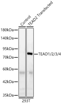
Western blot analysis of lysates from wild type (WT) and 293T cells transfected with TEAD2 using TEAD1/2/3/4 Rabbit mAb(CAB5092) at 1:1000 dilution incubated overnight at 4℃. Secondary antibody: HRP-conjugated Goat anti-Rabbit IgG (H+L) (CABS014) at 1:10000 dilution. Lysates/proteins: 25 μg per lane. Blocking buffer: 3% nonfat dry milk in TBST. Detection: ECL Basic Kit (AbGn00020). Exposure time: 20 s.
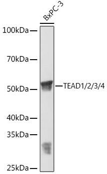
Western blot analysis of various lysates using TEAD1/2/3/4 Rabbit mAb (CAB5092) at 1:3000 dilution incubated overnight at 4℃. Secondary antibody: HRP-conjugated Goat anti-Rabbit IgG (H+L) (CABS014) at 1:10000 dilution. Lysates/proteins: 25μg per lane. Blocking buffer: 3% nonfat dry milk in TBST. Detection: ECL Basic Kit (AbGn00020). Exposure time: 1 min.
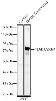
Western blot analysis of lysates from wild type (WT) and 293T cells transfected with TEAD4 using TEAD1/2/3/4 Rabbit mAb(CAB5092) at 1:1000 dilution incubated overnight at 4℃. Secondary antibody: HRP-conjugated Goat anti-Rabbit IgG (H+L) (CABS014) at 1:10000 dilution. Lysates/proteins: 25 μg per lane. Blocking buffer: 3% nonfat dry milk in TBST. Detection: ECL Basic Kit (AbGn00020). Exposure time: 10 s.
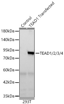
Western blot analysis of lysates from wild type (WT) and 293T cells transfected with TEAD1 using TEAD1/2/3/4 Rabbit mAb (CAB5092) at 1:1000 dilution incubated overnight at 4℃. Secondary antibody: HRP-conjugated Goat anti-Rabbit IgG (H+L) (CABS014) at 1:10000 dilution. Lysates/proteins: 25 μg per lane. Blocking buffer: 3% nonfat dry milk in TBST. Detection: ECL Basic Kit (AbGn00020) .Exposure time: 10 s.