The TEAD1 Monoclonal Antibody (CAB5092) is a high-quality antibody developed for reliable detection and analysis of target proteins. This gene encodes a ubiquitous transcriptional enhancer factor that is a member of the TEA/ATTS domain family. This protein directs the transactivation of a wide variety of genes and, in placental cells, also acts as a transcriptional repressor. Mutations in this gene cause Sveinsson's chorioretinal atrophy. Additional transcript variants have been described but their full-length natures have not been experimentally verified.
This antibody is validated for use in WB, ELISA applications and has demonstrated reactivity against Human samples.
Product Name:
TEAD1 Monoclonal Antibody
SKU:
CAB5092
Size:
100μL, 20μL
Reactivity:
Human
Clone Number:
ARC1158
Conjugate:
Unconjugated
Immunogen:
Recombinant protein (or fragment).This information is considered to be commercially sensitive.
Tested Applications:
WBELISA
Recommended Dilution:
WB
1:1000 - 1:5000
ELISA
Recommended starting concentration is 1 μg/mL. Please optimize the concentration based on your specific assay requirements.
Synonyms:
AA, REF1, TCF13, TEF-1, NTEF-1, TCF-13, TEAD-1
Positive Sample:
BXPC-3, 293T transfected with TEAD2 (Human), 293F transfected with TEAD3 (Human), 293T transfected with TEAD4 (Human), 293T transfected with TEAD1 (Human)
Cellular Localization:
Nucleus.
Calculated MW:
48 kDa/49 kDa/47 kDa
Observed MW:
53 kDa/90 kDa (Full-length protein)
This gene encodes a ubiquitous transcriptional enhancer factor that is a member of the TEA/ATTS domain family. This protein directs the transactivation of a wide variety of genes and, in placental cells, also acts as a transcriptional repressor. Mutations in this gene cause Sveinsson's chorioretinal atrophy. Additional transcript variants have been described but their full-length natures have not been experimentally verified.
Purification Method
Affinity purification
Gene ID
7003 7004 8463 7005
RRID
AB_2863440
Buffer Information
Store at -20℃. Avoid freeze / thaw cycles. Buffer: PBS containing 50% glycerol and 0.05% BSA, preserved with proclin300 or sodium azide, pH 7.3.
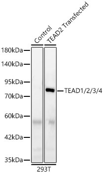
Western blot analysis of lysates from wild type (WT) and 293T cells transfected with TEAD2 using TEAD1/2/3/4 Rabbit mAb(CAB5092) at 1:1000 dilution incubated overnight at 4℃. Secondary antibody: HRP-conjugated Goat anti-Rabbit IgG (H+L) (AS014) at 1:10000 dilution. Lysates/proteins: 25 μg per lane. Blocking buffer: 3% nonfat dry milk in TBST. Detection: ECL Basic Kit (AbGn00020). Exposure time: 20 s.
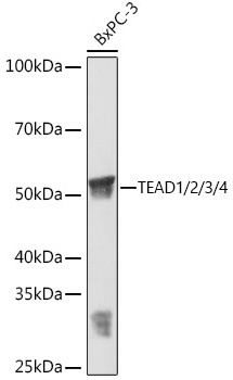
Western blot analysis of various lysates using TEAD1/2/3/4 Rabbit mAb (CAB5092) at 1:3000 dilution incubated overnight at 4℃. Secondary antibody: HRP-conjugated Goat anti-Rabbit IgG (H+L) (AS014) at 1:10000 dilution. Lysates/proteins: 25μg per lane. Blocking buffer: 3% nonfat dry milk in TBST. Detection: ECL Basic Kit (AbGn00020). Exposure time: 1 min.
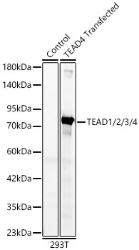
Western blot analysis of lysates from wild type (WT) and 293T cells transfected with TEAD4 using TEAD1/2/3/4 Rabbit mAb(CAB5092) at 1:1000 dilution incubated overnight at 4℃. Secondary antibody: HRP-conjugated Goat anti-Rabbit IgG (H+L) (AS014) at 1:10000 dilution. Lysates/proteins: 25 μg per lane. Blocking buffer: 3% nonfat dry milk in TBST. Detection: ECL Basic Kit (AbGn00020). Exposure time: 10 s.
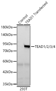
Western blot analysis of lysates from wild type (WT) and 293T cells transfected with TEAD1 using TEAD1/2/3/4 Rabbit mAb (CAB5092) at 1:1000 dilution incubated overnight at 4℃. Secondary antibody: HRP-conjugated Goat anti-Rabbit IgG (H+L) (AS014) at 1:10000 dilution. Lysates/proteins: 25 μg per lane. Blocking buffer: 3% nonfat dry milk in TBST. Detection: ECL Basic Kit (AbGn00020) .Exposure time: 10 s.