The TEAD1 Antibody (CAB6768) is a high-quality antibody developed for reliable detection and analysis of target proteins. This antibody is produced in rabbits and has a high specificity for human samples, making it an excellent choice for Western blot applications. By binding to TEAD1, this antibody enables precise detection and analysis of TEAD1 protein levels in various cell types.TEAD1 is a key player in regulating gene expression and cell proliferation, making it a critical target for studies in developmental biology, cancer research, and regenerative medicine.
This antibody is validated for use in WB, IHC-P, IF/ICC, IP, ChIP, ELISA applications and has demonstrated reactivity against Human, Mouse, Rat samples.
Product Name:
TEAD1 Antibody
SKU:
CAB6768
Size:
20μL, 100μL
Reactivity:
Human, Mouse, Rat
Conjugate:
Unconjugated
Immunogen:
Recombinant protein (or fragment).This information is considered to be commercially sensitive.
Sequence:
AIHN KLGL PGIP RPTF PGAP GFWP GMIQ TGQP GSSQ DVKP FVQQ AYPI QPAV TAPI PGFE PASA PAPS VPAW QGRS IGTT K
Tested Applications:
WBIHC-PIF/ICCIPChIPELISA
Recommended Dilution:
WB
1:1000 - 1:5000
IHC-P
1:50 - 1:200
IF/ICC
1:50 - 1:200
IP
0.5μg-4μg antibody for 400μg-600μg extracts of whole cells
ELISA
Recommended starting concentration is 1 μg/mL. Please optimize the concentration based on your specific assay requirements.
This gene encodes a ubiquitous transcriptional enhancer factor that is a member of the TEA/ATTS domain family. This protein directs the transactivation of a wide variety of genes and, in placental cells, also acts as a transcriptional repressor. Mutations in this gene cause Sveinsson's chorioretinal atrophy. Additional transcript variants have been described but their full-length natures have not been experimentally verified.
Purification Method
Affinity purification
Gene ID
7003
RRID
AB_2767351
Buffer Information
Store at -20℃. Avoid freeze / thaw cycles. Buffer: PBS containing 50% glycerol, preserved with proclin300 or sodium azide, pH 7.3.
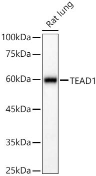
Western blot analysis of lysates from Rat lung using TEAD1 Rabbit pAb (CAB6768) at 1:2000 dilution. Secondary antibody: HRP-conjugated Goat anti-Rabbit IgG (H+L) (CABS014) at 1:10000 dilution. Lysates/proteins: 25 μg per lane. Blocking buffer: 3% nonfat dry milk in TBST. Detection: ECL Basic Kit (AbGn00020). Exposure time:90s.
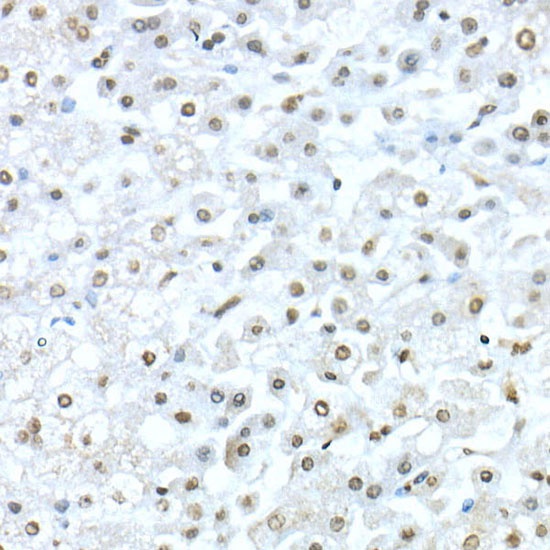
Immunohistochemistry analysis of paraffin-embedded Human liver using TEAD1 Rabbit pAb (CAB6768) at dilution of 1:50 (40x lens). High pressure antigen retrieval performed with 0.01M Citrate buffer (pH 6.0) prior to IHC staining.
Immunohistochemistry analysis of paraffin-embedded Human liver using TEAD1 Rabbit pAb (CAB6768) at dilution of 1:50 (40x lens). High pressure antigen retrieval performed with 0.01M Citrate buffer (pH 6.0) prior to IHC staining.
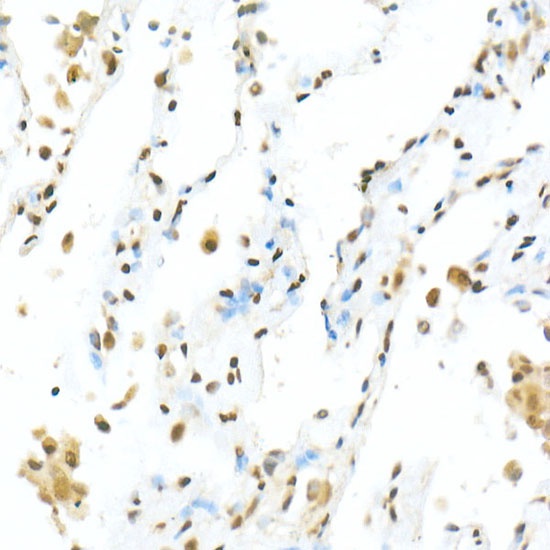
Immunohistochemistry analysis of paraffin-embedded Human lung using TEAD1 Rabbit pAb (CAB6768) at dilution of 1:50 (40x lens). High pressure antigen retrieval performed with 0.01M Citrate buffer (pH 6.0) prior to IHC staining.
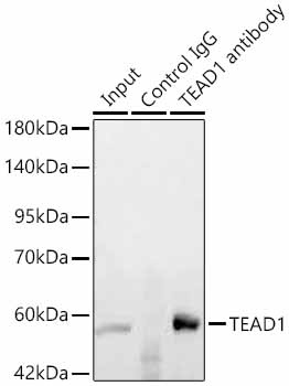
Immunoprecipitation of TEAD1 in 600 µg extracts from mouse heart using 3 µg TEAD1 Rabbit pAb (CAB6768). Western blot analysis was performed using TEAD1 Rabbit pAb (CAB6768) at 1:200 dilution.
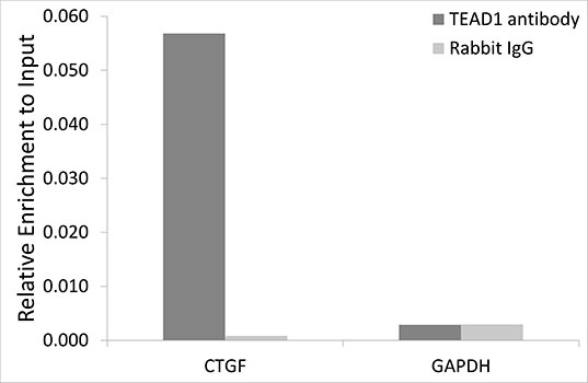
Chromatin immunoprecipitation analysis of extracts of HeLa cells, using TEAD1 antibody (CAB6768) and rabbit IgG. The amount of immunoprecipitated DNA was checked by quantitative PCR. Histogram was constructed by the ratios of the immunoprecipitated DNA to the input.