The TGF beta induced TGFBI Monoclonal Antibody (CAB2407) is a high-quality antibody developed for reliable detection and analysis of target proteins. This antibody, developed using rabbit monoclonal technology, offers high specificity and sensitivity in detecting TGFBI in human samples, making it a reliable choice for Western blot experiments.TGFBI, also known as BIGH3, has been implicated in various diseases, including corneal dystrophies, cancer metastasis, and fibrotic disorders. By targeting TGFBI with this antibody, researchers can gain valuable insights into the mechanisms underlying these conditions and potentially identify new therapeutic targets.
This antibody is validated for use in WB, IHC-P, ELISA applications and has demonstrated reactivity against Human, Mouse, Rat samples.
Product Name:
TGF beta induced TGFBI Monoclonal Antibody
SKU:
CAB2407
Size:
20μL, 100μL
Reactivity:
Human, Mouse, Rat
Clone Number:
ARC0757
Conjugate:
Unconjugated
Immunogen:
Synthetic peptide. This information is considered to be commercially sensitive.
This gene encodes an RGD-containing protein that binds to type I, II and IV collagens. The RGD motif is found in many extracellular matrix proteins modulating cell adhesion and serves as a ligand recognition sequence for several integrins. This protein plays a role in cell-collagen interactions and may be involved in endochondrial bone formation in cartilage. The protein is induced by transforming growth factor-beta and acts to inhibit cell adhesion. Mutations in this gene are associated with multiple types of corneal dystrophy.
Purification Method
Affinity purification
Gene ID
7045
RRID
AB_2863002
Buffer Information
Store at -20℃. Avoid freeze / thaw cycles. Buffer: PBS containing 50% glycerol and 0.05% BSA, preserved with proclin300 or sodium azide, pH 7.3.
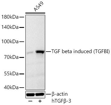
Western blot analysis of lysates from A549 cells using TGF beta induced (TGFBI) Rabbit mAb (CAB2407) at 1:1000 dilution incubated overnight at 4℃. A549 treated with hTGFβ-3 (100 ng/ml) at 37℃ for 16 hours. Secondary antibody: HRP-conjugated Goat anti-Rabbit IgG (H+L) (CABS014) at 1:10000 dilution. Lysates/proteins: 30 μg per lane. Blocking buffer: 3 % nonfat dry milk in TBST. Detection: ECL Basic Kit (AbGn00020). Exposure time: 45 s.
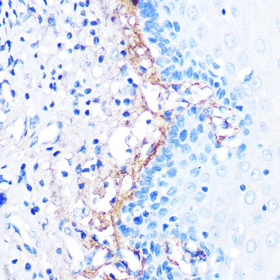
Immunohistochemistry analysis of paraffin-embedded Human esophageal using TGF beta induced TGF beta induced (TGFBI) Rabbit mAb (CAB2407) at dilution of 1:100 (40x lens). Microwave antigen retrieval performed with 0.01M PBS Buffer (pH 7.2) prior to IHC staining.