The THAP2 Antibody (CAB3518) is a high-quality antibody developed for reliable detection and analysis of target proteins. This antibody, derived from rabbits, exhibits high reactivity with human samples and is validated for use in Western blot applications.THAP2 is a member of the THAP protein family, known for its DNA-binding capabilities and its role in gene regulation. The THAP2 protein has been implicated in the development and progression of cancer, making it a promising target for cancer research.
This antibody is validated for use in WB, ELISA applications and has demonstrated reactivity against Mouse, Rat samples.
Product Name:
THAP2 Antibody
SKU:
CAB3518
Size:
20μL, 100μL
Reactivity:
Mouse, Rat
Conjugate:
Unconjugated
Immunogen:
Recombinant protein (or fragment).This information is considered to be commercially sensitive.
Recommended starting concentration is 1 μg/mL. Please optimize the concentration based on your specific assay requirements.
Synonyms:
THAP2
Positive Sample:
Mouse small intestine, Rat brain
Cellular Localization:
Nucleolus, Nucleus.
Calculated MW:
26kDa
Observed MW:
30kDa
Predicted to enable DNA binding activity and metal ion binding activity. Located in nucleolus.
Purification Method
Affinity purification
Gene ID
83591
RRID
AB_2765145
Buffer Information
Store at -20℃. Avoid freeze / thaw cycles. Buffer: PBS with 0.01% thimerosal,50% glycerol,pH7.3.
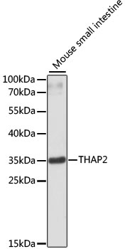
Western blot analysis of lysates from mouse small intestine, using THAP2 Rabbit pAb (CAB3518) at 1:3000 dilution. Secondary antibody: HRP-conjugated Goat anti-Rabbit IgG (H+L) (CABS014) at 1:10000 dilution. Lysates/proteins: 25μg per lane. Blocking buffer: 3% nonfat dry milk in TBST. Detection: ECL Basic Kit (AbGn00020). Exposure time: 90s.
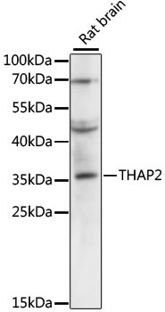
Western blot analysis of lysates from rat brain, using THAP2 Rabbit pAb (CAB3518) at 1:3000 dilution. Secondary antibody: HRP-conjugated Goat anti-Rabbit IgG (H+L) (CABS014) at 1:10000 dilution. Lysates/proteins: 25μg per lane. Blocking buffer: 3% nonfat dry milk in TBST. Detection: ECL Enhanced Kit (AbGn00021). Exposure time: 5s.