The TNFAIP8L2 Antibody (CAB13698) is a high-quality antibody developed for reliable detection and analysis of target proteins. This antibody, raised in rabbits, is highly specific for human samples and has been validated for use in Western blot applications. By binding to the TNFAIP8L2 protein, the antibody allows for the detection and analysis of TNFAIP8L2 in various cell types, making it ideal for studies in immunology and cancer research.
This antibody is validated for use in WB, IF/ICC, IP, ELISA applications and has demonstrated reactivity against Human, Mouse, Rat samples.
Product Name:
TNFAIP8L2 Antibody
SKU:
CAB13698
Size:
20μL, 100μL
Reactivity:
Human, Mouse, Rat
Conjugate:
Unconjugated
Immunogen:
Recombinant protein (or fragment).This information is considered to be commercially sensitive.
0.5μg-4μg antibody for 25μg-100μg extracts of whole cells
ELISA
Recommended starting concentration is 1 μg/mL. Please optimize the concentration based on your specific assay requirements.
Synonyms:
TIPE2, TNFAIP8L2
Positive Sample:
THP-1, Mouse thymus, Mouse spleen, Rat spleen
Cellular Localization:
Cytoplasm.
Calculated MW:
21kDa
Observed MW:
18kDa
Predicted to be involved in negative regulation of T cell activation and negative regulation of inflammatory response. Predicted to be active in cytoplasm.
Purification Method
Affinity purification
Gene ID
79626
RRID
AB_2760558
Buffer Information
Store at -20℃. Avoid freeze / thaw cycles. Buffer: PBS with 0.01% thimerosal,50% glycerol,pH7.3.
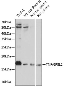
Western blot analysis of various lysates using TNFAIP8L2 Rabbit pAb (CAB13698) at 1:1000 dilution. Secondary antibody: HRP-conjugated Goat anti-Rabbit IgG (H+L) (CABS014) at 1:10000 dilution. Lysates/proteins: 25μg per lane. Blocking buffer: 3% nonfat dry milk in TBST. Detection: ECL Basic Kit (AbGn00020). Exposure time: 10s.
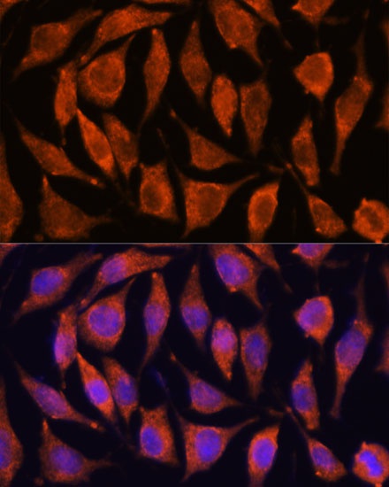
Immunofluorescence analysis of C6 cells using TNFAIP8L2 Rabbit pAb (CAB13698) at dilution of 1:100. Secondary antibody: Cy3-conjugated Goat anti-Rabbit IgG (H+L) (CABS007) at 1:500 dilution. Blue: DAPI for nuclear staining.
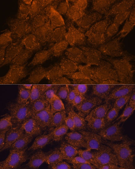
Immunofluorescence analysis of L929 cells using TNFAIP8L2 Rabbit pAb (CAB13698) at dilution of 1:100. Secondary antibody: Cy3-conjugated Goat anti-Rabbit IgG (H+L) (CABS007) at 1:500 dilution. Blue: DAPI for nuclear staining.
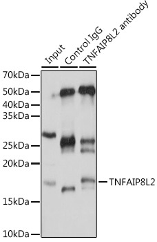
Western blot analysis of various lysates using TNFAIP8L2 Rabbit pAb (CAB13698) at 1:1000 dilution. Secondary antibody: HRP-conjugated Goat anti-Rabbit IgG (H+L) (CABS014) at 1:10000 dilution. Lysates/proteins: 25μg per lane. Blocking buffer: 3% nonfat dry milk in TBST. Detection: ECL Basic Kit (AbGn00020). Exposure time: 10s.