The TSC2 Antibody (CAB0492) is a high-quality antibody developed for reliable detection and analysis of target proteins. This antibody, generated in rabbits, is highly specific to human samples and has been validated for use in Western blot applications. By targeting the TSC2 protein, researchers can detect and analyze its expression in various cell types, making it a versatile tool for studies in molecular biology and genetic disorders.TSC2 is a critical regulator of cell growth and proliferation, with mutations in the gene leading to the development of tumors and other manifestations of tuberous sclerosis complex.
This antibody is validated for use in WB, IHC-P, IF/ICC, ELISA applications and has demonstrated reactivity against Human, Mouse, Rat samples.
Product Name:
TSC2 Antibody
SKU:
CAB0492
Size:
20μL, 100μL
Reactivity:
Human, Mouse, Rat
Conjugate:
Unconjugated
Immunogen:
Recombinant protein (or fragment).This information is considered to be commercially sensitive.
Recommended starting concentration is 1 μg/mL. Please optimize the concentration based on your specific assay requirements.
Synonyms:
LAM, TSC4, PPP1R160, TSC2
Positive Sample:
SH-SY5Y
Cellular Localization:
Cytoplasm, Membrane, Peripheral Membrane Protein.
Calculated MW:
201kDa
Observed MW:
200kDa
This gene is a tumor suppressor gene that encodes the growth inhibitory protein tuberin. Tuberin interacts with hamartin to form the TSC protein complex which functions in the control of cell growth. This TSC protein complex negatively regulates mammalian target of rapamycin complex 1 (mTORC1) signaling which is a major regulator of anabolic cell growth. Mutations in this gene have been associated with tuberous sclerosis and lymphangioleiomyomatosis.
Purification Method
Affinity purification
Gene ID
7249
RRID
AB_2757221
Buffer Information
Store at -20℃. Avoid freeze / thaw cycles. Buffer: PBS containing 50% glycerol, preserved with proclin300 or sodium azide, pH 7.3.
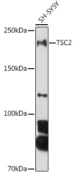
Western blot analysis of lysates from SH-SY5Y cells, using TSC2 Rabbit pAb (CAB0492) at 1:1000 dilution. Secondary antibody: HRP-conjugated Goat anti-Rabbit IgG (H+L) (CABS014) at 1:10000 dilution. Lysates/proteins: 25μg per lane. Blocking buffer: 3% nonfat dry milk in TBST. Detection: ECL Basic Kit (AbGn00020). Exposure time: 180s.
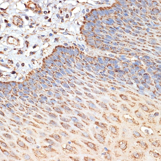
Immunohistochemistry analysis of paraffin-embedded Human esophageal using TSC2 Rabbit pAb (CAB0492) at dilution of 1:100 (40x lens). Microwave antigen retrieval performed with 0.01M Tris/EDTA Buffer (pH 9.0) prior to IHC staining.