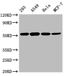
| Western Blot Positive WB detected in: 293 whole cell lysate, A549 whole cell lysate, Hela whole cell lysate, MCF-7 whole cell lysate All lanes TUBB antibody at 1:5000 Secondary Goat polyclonal to mouse IgG at 1/50000 dilution Predicted band size: 55 KDa Observed band size: 55 KDa Exposure time:5s |
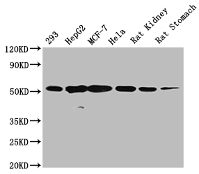
 | Western Blot Positive WB detected in: 293 whole cell lysate, HepG2 whole cell lysate, MCF-7 whole cell lysate, Hela whole cell lysate, Rat kidney tissue, Rat stomach tissue All lanes TUBB antibody at 1:5000 Secondary Goat polyclonal to mouse IgG at 1/50000 dilution Predicted band size: 55 KDa Observed band size: 55 KDa Exposure time:5min |
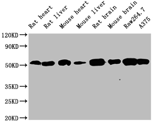 | Western Blot Positive WB detected in: Rat heart tissue,Rat liver tissue, Mouse heart tissue, Mouse liver tissue, Rat brain tissue, Mouse brain tissue, Raw264.7 whole cell lysate, A375 whole cell lysate All lanes TUBB antibody at 1:5000 Secondary Goat polyclonal to mouse IgG at 1/50000 dilution Predicted band size: 55 KDa Observed band size: 55 KDa Exposure time:5min |
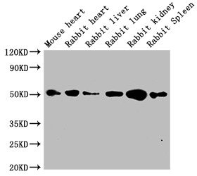
 | Western Blot Positive WB detected in: Mouse heart tissue, Rabbit heart tissue, Rabbit liver tissue, Rabbit lung tissue, Rabbit kidney tissue, Rabbit spleen tissue All lanes TUBB antibody at 1:5000 Secondary Goat polyclonal to mouse IgG at 1/50000 dilution Predicted band size: 55 KDa Observed band size: 55 KDa Exposure time:5min |
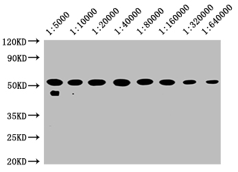
 | Western Blot Positive WB detected in: 20µg hela whole cell lysate TUBB antibody at 1:5000, 1:10000, 1:20000, 1:40000, 1:80000, 1:160000, 1:320000, 1:640000 Secondary Goat polyclonal to mouse IgG at 1/50000 dilution Predicted band size: 55 KDa Observed band size: 55 KDa Exposure time:5min |
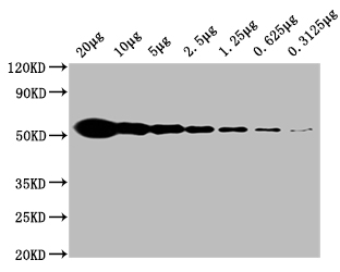 | Western Blot Positive WB detected in: Hela whole cell lysate at 20µg, 10µg, 5µg, 2.5µg, 1.25µg, 0.625µg, 0.3125µg All lanes:TUBB antibody at 1:5000 Secondary Goat polyclonal to mouse IgG at 1/50000 dilution Predicted band size: 55 KDa Observed band size: 55 KDa Exposure time:5min |
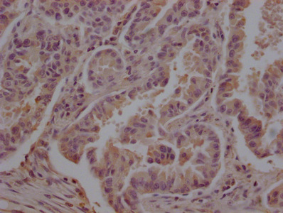 | IHC image of MACO0666 diluted at 1:200 and staining in paraffin-embedded human lung cancer performed on a Leica BondTM system. After dewaxing and hydration, antigen retrieval was mediated by high pressure in a citrate buffer (pH 6.0). Section was blocked with 10% normal goat serum 30min at 37°C Then primary antibody (1% BSA) was incubated at 4°C overnight. The primary is detected by a Goat anti-Mouse IgG labeled by HRP and visualized using 0.05% DAB. |
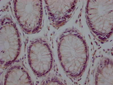
 | IHC image of MACO0666 diluted at 1:200 and staining in paraffin-embedded human colon cancer performed on a Leica BondTM system. After dewaxing and hydration, antigen retrieval was mediated by high pressure in a citrate buffer (pH 6.0). Section was blocked with 10% normal goat serum 30min at 37°C Then primary antibody (1% BSA) was incubated at 4°C overnight. The primary is detected by a Goat anti-Mouse IgG labeled by HRP and visualized using 0.05% DAB. |
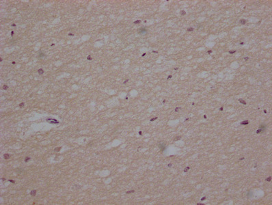 | IHC image of MACO0666 diluted at 1:200 and staining in paraffin-embedded human brain tissue performed on a Leica BondTM system. After dewaxing and hydration, antigen retrieval was mediated by high pressure in a citrate buffer (pH 6.0). Section was blocked with 10% normal goat serum 30min at 37°C Then primary antibody (1% BSA) was incubated at 4°C overnight. The primary is detected by a Goat anti-Mouse IgG labeled by HRP and visualized using 0.05% DAB. |
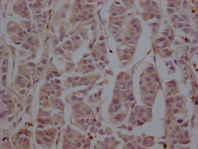
 | IHC image of MACO0666 diluted at 1:200 and staining in paraffin-embedded human breast cancer performed on a Leica BondTM system. After dewaxing and hydration, antigen retrieval was mediated by high pressure in a citrate buffer (pH 6.0). Section was blocked with 10% normal goat serum 30min at 37°C Then primary antibody (1% BSA) was incubated at 4°C overnight. The primary is detected by a Goat anti-Mouse IgG labeled by HRP and visualized using 0.05% DAB. |
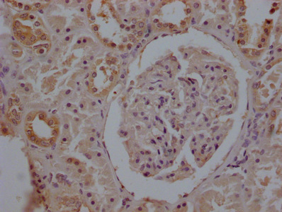 | IHC image of MACO0666 diluted at 1:200 and staining in paraffin-embedded human kidney tissue performed on a Leica BondTM system. After dewaxing and hydration, antigen retrieval was mediated by high pressure in a citrate buffer (pH 6.0). Section was blocked with 10% normal goat serum 30min at 37°C Then primary antibody (1% BSA) was incubated at 4°C overnight. The primary is detected by a Goat anti-Mouse IgG labeled by HRP and visualized using 0.05% DAB. |
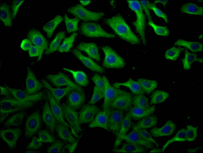
 | Immunofluorescence staining of NIH/3T3 cells with MACO0666 at 1:100, counter-stained with DAPI. The cells were fixed in 4% formaldehyde, permeated by 0.2% TritonX-100, and blocked in 10% normal Goat Serum. The cells were then incubated with the antibody overnight at 4°C. Nuclear DNA was labeled in blue with DAPI. The secondary antibody was FITC-conjugated AffiniPure Goat Anti-Mouse IgG(H+L). |
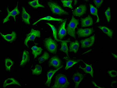
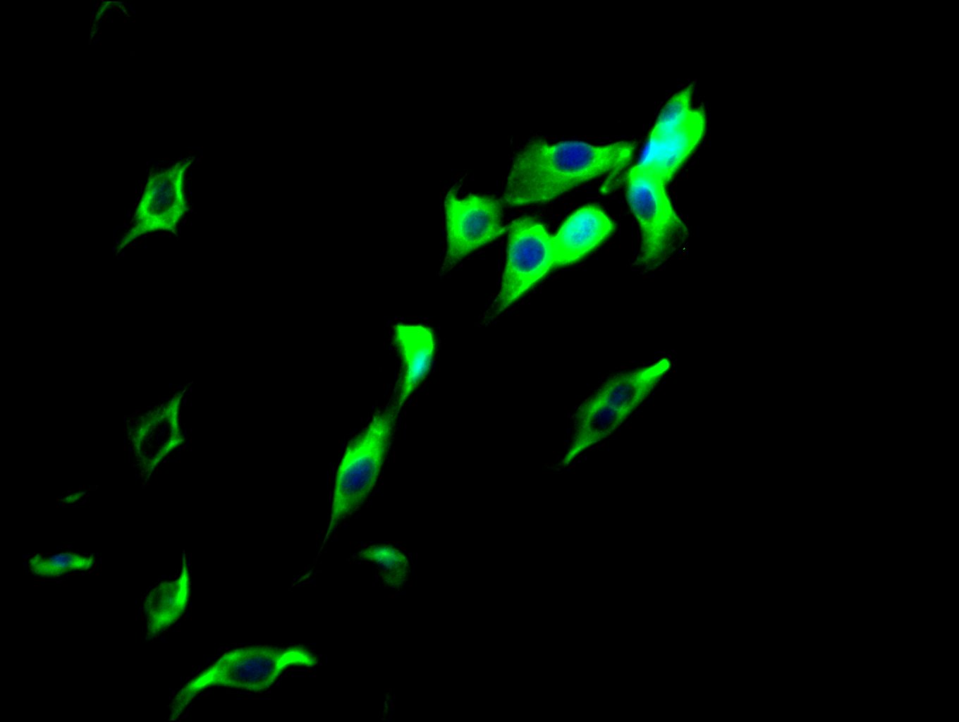
 | Immunofluorescence staining of A549 cells with MACO0666 at 1:100, counter-stained with DAPI. The cells were fixed in 4% formaldehyde, permeated by 0.2% TritonX-100, and blocked in 10% normal Goat Serum. The cells were then incubated with the antibody overnight at 4°C. Nuclear DNA was labeled in blue with DAPI. The secondary antibody was FITC-conjugated AffiniPure Goat Anti-Mouse IgG(H+L). |
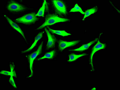
 | Immunofluorescence staining of Hela cells with MACO0666 at 1:100, counter-stained with DAPI. The cells were fixed in 4% formaldehyde, permeated by 0.2% TritonX-100, and blocked in 10% normal Goat Serum. The cells were then incubated with the antibody overnight at 4°C. Nuclear DNA was labeled in blue with DAPI. The secondary antibody was FITC-conjugated AffiniPure Goat Anti-Mouse IgG(H+L). |
 | Immunofluorescence staining of HepG2 cells with MACO0666 at 1:100, counter-stained with DAPI. The cells were fixed in 4% formaldehyde, permeated by 0.2% TritonX-100, and blocked in 10% normal Goat Serum. The cells were then incubated with the antibody overnight at 4°C. Nuclear DNA was labeled in blue with DAPI. The secondary antibody was FITC-conjugated AffiniPure Goat Anti-Mouse IgG(H+L). |
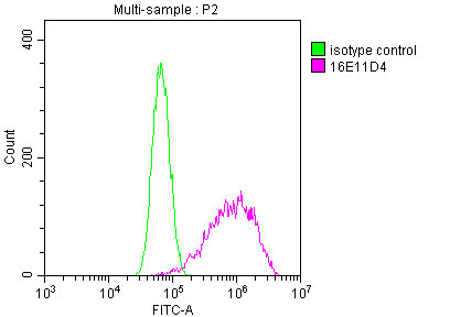
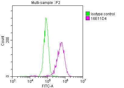
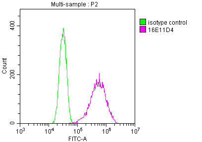
 | Overlay Peak curve showing A549 cells stained with MACO0666 (red line) at 1:200. The cells were fixed in 4% formaldehyde and permeated by 0.2% TritonX-100. Then 10% normal goat serum was Incubated to block non-specific protein-protein interactions followed by the antibody (1µg/1*106cells) for 1 h at 4°C. The secondary antibody used was FITC-conjugated Goat Anti-Mouse IgG(H+L) at 1/100 dilution for 30min at 4°C. Isotype control antibody (green line) was mouse IgG2b (1µg/1*106cells) used under the same conditions. Acquisition of >10,000 events was performed. |
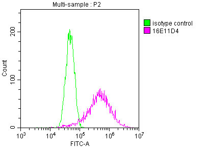
 | Overlay Peak curve showing HepG2 cells stained with MACO0666 (red line) at 1:200. The cells were fixed in 4% formaldehyde and permeated by 0.2% TritonX-100. Then 10% normal goat serum was Incubated to block non-specific protein-protein interactions followed by the antibody (1µg/1*106cells) for 1 h at 4°C. The secondary antibody used was FITC-conjugated Goat Anti-Mouse IgG(H+L) at 1/100 dilution for 30min at 4°C. Isotype control antibody (green line) was mouse IgG2b (1µg/1*106cells) used under the same conditions. Acquisition of >10,000 events was performed. |
 | Overlay Peak curve showing MCF-7 cells stained with MACO0666 (red line) at 1:200. The cells were fixed in 4% formaldehyde and permeated by 0.2% TritonX-100. Then 10% normal goat serum was Incubated to block non-specific protein-protein interactions followed by the antibody (1µg/1*106cells) for 1 h at 4°C. The secondary antibody used was FITC-conjugated Goat Anti-Mouse IgG(H+L) at 1/100 dilution for 30min at 4°C. Isotype control antibody (green line) was mouse IgG2b (1µg/1*106cells) used under the same conditions. Acquisition of >10,000 events was performed. |
 | Overlay Peak curve showing NIH/3T3 cells stained with MACO0666 (red line) at 1:200. The cells were fixed in 4% formaldehyde and permeated by 0.2% TritonX-100. Then 10% normal goat serum was Incubated to block non-specific protein-protein interactions followed by the antibody (1µg/1*106cells) for 1 h at 4°C. The secondary antibody used was FITC-conjugated Goat Anti-Mouse IgG(H+L) at 1/100 dilution for 30min at 4°C. Isotype control antibody (green line) was mouse IgG2b (1µg/1*106cells) used under the same conditions. Acquisition of >10,000 events was performed. |
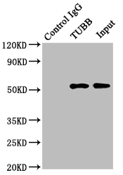 | Immunoprecipitating TUBB in Hela whole cell lysate Lane 1: Mouse control IgG instead of MACO0666 in Hela whole cell lysate. Lane 2: MACO0666 (2µg) + Hela whole cell lysate (500µg) Lane 3: Hela whole cell lysate (5µg) For western blotting, the blot was detected with MACO0666 at 1:2000, and a HRP-conjugated Protein G antibody was used as the secondary antibody at 1:5000 |