The UBE2B Monoclonal Antibody (CAB0503) is a high-quality antibody developed for reliable detection and analysis of target proteins. This antibody, generated in rabbits, exhibits high reactivity with human samples and has been validated for use in Western blot applications. By binding to the UBE2B protein, researchers can study its role in various cellular processes, making it an essential tool for studies in molecular biology and cancer research.UBE2B plays a crucial role in the ubiquitin-proteasome system, a pathway responsible for protein degradation and regulation.
This antibody is validated for use in WB, IHC-P, ELISA applications and has demonstrated reactivity against Human, Mouse, Rat samples.
Product Name:
UBE2B Monoclonal Antibody
SKU:
CAB0503
Size:
20μL, 100μL
Reactivity:
Human, Mouse, Rat
Clone Number:
ARC2512
Conjugate:
Unconjugated
Immunogen:
Synthetic peptide. This information is considered to be commercially sensitive.
Recommended starting concentration is 1 μg/mL. Please optimize the concentration based on your specific assay requirements.
Synonyms:
HR6B, UBC2, HHR6B, RAD6B, E2-17kDa, UBE2B
Positive Sample:
HeLa, 293T, NIH/3T3, K-562, Mouse testis, Mouse heart, Rat testis
Cellular Localization:
Cell Membrane, Nucleus.
Calculated MW:
17kDa
Observed MW:
17kDa
The modification of proteins with ubiquitin is an important cellular mechanism for targeting abnormal or short-lived proteins for degradation. Ubiquitination involves at least three classes of enzymes: ubiquitin-activating enzymes, or E1s, ubiquitin-conjugating enzymes, or E2s, and ubiquitin-protein ligases, or E3s. This gene encodes a member of the E2 ubiquitin-conjugating enzyme family. This enzyme is required for post-replicative DNA damage repair. Its protein sequence is 100% identical to the mouse, rat, and rabbit homologs, which indicates that this enzyme is highly conserved in eukaryotic evolution.
Purification Method
Affinity purification
Gene ID
7320
Buffer Information
Store at -20℃. Avoid freeze / thaw cycles. Buffer: PBS containing 50% glycerol and 0.05% BSA, preserved with proclin300 or sodium azide, pH 7.3.
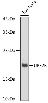
Western blot analysis of lysates from Rat testis, using UBE2B Rabbit mAb (CAB0503) at 1:1000 dilution. Secondary antibody: HRP-conjugated Goat anti-Rabbit IgG (H+L) (CABS014) at 1:10000 dilution. Lysates/proteins: 25μg per lane. Blocking buffer: 3% nonfat dry milk in TBST. Detection: ECL Basic Kit (AbGn00020). Exposure time: 1s.
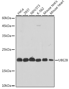
Western blot analysis of various lysates using UBE2B Rabbit mAb (CAB0503) at 1:1000 dilution. Secondary antibody: HRP-conjugated Goat anti-Rabbit IgG (H+L) (CABS014) at 1:10000 dilution. Lysates/proteins: 25μg per lane. Blocking buffer: 3% nonfat dry milk in TBST. Detection: ECL Basic Kit (AbGn00020). Exposure time: 3s.
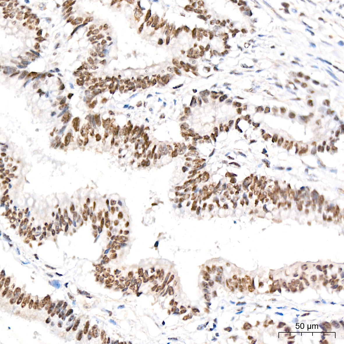
Immunohistochemistry analysis of paraffin-embedded Human colon carcinoma tissue using UBE2B Rabbit mAb (CAB0503) at a dilution of 1:800 (40x lens). High pressure antigen retrieval performed with 0.01M Citrate buffer (pH 6.0) prior to IHC staining.
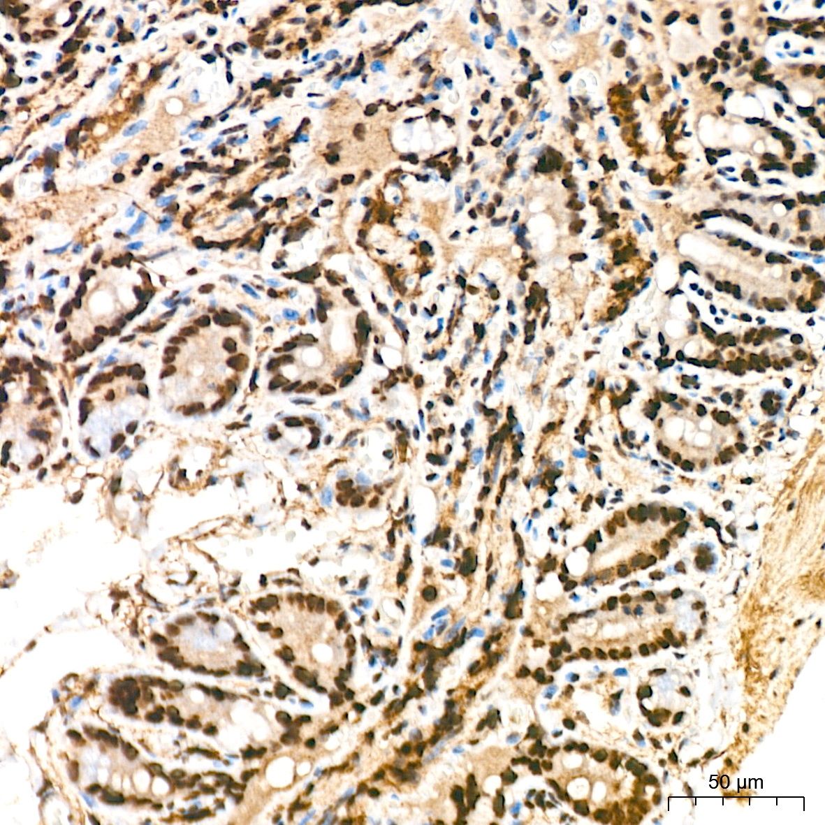
Immunohistochemistry analysis of paraffin-embedded Mouse colon tissue using UBE2B Rabbit mAb (CAB0503) at a dilution of 1:800 (40x lens). High pressure antigen retrieval performed with 0.01M Citrate buffer (pH 6.0) prior to IHC staining.
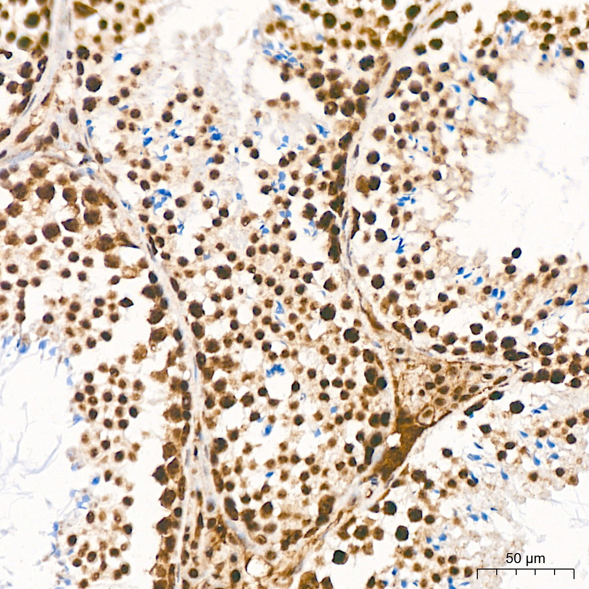
Immunohistochemistry analysis of paraffin-embedded Mouse testis tissue using UBE2B Rabbit mAb (CAB0503) at a dilution of 1:800 (40x lens). High pressure antigen retrieval performed with 0.01M Citrate buffer (pH 6.0) prior to IHC staining.
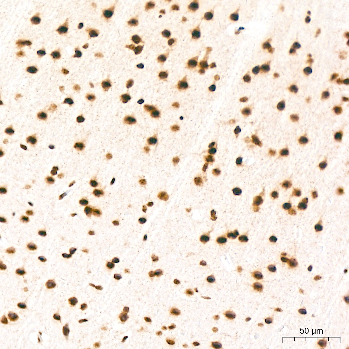
Immunohistochemistry analysis of paraffin-embedded Rat brain tissue using UBE2B Rabbit mAb (CAB0503) at a dilution of 1:800 (40x lens). High pressure antigen retrieval performed with 0.01M Citrate buffer (pH 6.0) prior to IHC staining.