The ULK1 Antibody (CAB8529) is a high-quality antibody developed for reliable detection and analysis of target proteins. This antibody, produced in rabbits, exhibits high reactivity with human samples and is suitable for use in Western blot applications. By binding specifically to the ULK1 protein, this antibody enables precise detection and analysis in various cell types, making it an essential component of studies in cell biology and disease research.ULK1, a serine/threonine kinase, plays a crucial role in autophagy initiation and maintenance by initiating the formation of autophagosomes.
This antibody is validated for use in WB, IF/ICC, ELISA applications and has demonstrated reactivity against Human, Mouse, Rat samples.
Product Name:
ULK1 Antibody
SKU:
CAB8529
Size:
20μL, 100μL
Reactivity:
Human, Mouse, Rat
Conjugate:
Unconjugated
Immunogen:
Synthetic peptide. This information is considered to be commercially sensitive.
Recommended starting concentration is 1 μg/mL. Please optimize the concentration based on your specific assay requirements.
Synonyms:
ATG1, ATG1A, UNC51, hATG1, Unc51.1, ULK1
Positive Sample:
293T, HepG2, Rat brain, Mouse thymus, Rat testis
Cellular Localization:
Cytoplasm, Preautophagosomal Structure, Cytosol.
Calculated MW:
113kDa
Observed MW:
150kDa/140kDa
Enables identical protein binding activity; protein serine/threonine kinase activity; and small GTPase binding activity. Involved in several processes, including autophagosome assembly; positive regulation by symbiont of host autophagy; and protein phosphorylation. Located in autophagosome; cytosol; and phagophore assembly site membrane. Is extrinsic component of autophagosome membrane; extrinsic component of omegasome membrane; and extrinsic component of phagophore assembly site membrane. Part of Atg1/ULK1 kinase complex.
Purification Method
Affinity purification
Gene ID
8408
RRID
AB_2772810
Buffer Information
Store at -20℃. Avoid freeze / thaw cycles. Buffer: PBS containing 50% glycerol, preserved with proclin300 or sodium azide, pH 7.3.
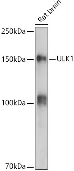
Western blot analysis of lysates from Rat brain, using ULK1 Rabbit pAb (CAB8529) at 1:500 dilution. Secondary antibody: HRP-conjugated Goat anti-Rabbit IgG (H+L) (CABS014) at 1:10000 dilution. Lysates/proteins: 25μg per lane. Blocking buffer: 3% nonfat dry milk in TBST. Detection: ECL Enhanced Kit (AbGn00021). Exposure time: 180s.
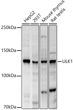
Western blot analysis of various lysates, using ULK1 Rabbit pAb (CAB8529) at 1:2000 dilution. Secondary antibody: HRP-conjugated Goat anti-Rabbit IgG (H+L) (CABS014) at 1:10000 dilution. Lysates/proteins: 25μg per lane. Blocking buffer: 3% nonfat dry milk in TBST. Detection: ECL Basic Kit (AbGn00020). Exposure time: 10s.
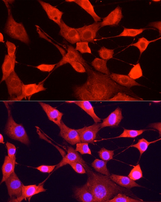
Immunofluorescence analysis of NIH/3T3 cells using ULK1 Rabbit pAb (CAB8529) at dilution of 1:200 (40x lens). Secondary antibody: Cy3-conjugated Goat anti-Rabbit IgG (H+L) (CABS007) at 1:500 dilution. Blue: DAPI for nuclear staining.


")