The UQCRC2 Monoclonal Antibody (CAB4366) is a high-quality antibody developed for reliable detection and analysis of target proteins. The protein encoded by this gene is located in the mitochondrion, where it is part of the ubiquinol-cytochrome c reductase complex (also known as complex III). This complex constitutes a part of the mitochondrial respiratory chain. Defects in this gene are a cause of mitochondrial complex III deficiency nuclear type 5.
This antibody is validated for use in WB, IHC-P, ELISA applications and has demonstrated reactivity against Human, Mouse, Rat samples.
Product Name:
UQCRC2 Monoclonal Antibody
SKU:
CAB4366
Size:
100μL, 20μL
Reactivity:
Human, Mouse, Rat
Clone Number:
ARC0982
Conjugate:
Unconjugated
Immunogen:
Synthetic peptide. This information is considered to be commercially sensitive.
Tested Applications:
WBIHC-PELISA
Recommended Dilution:
WB
1:1000 - 14000
IHC-P
1:500 - 1:2000
ELISA
Recommended starting concentration is 1 μg/mL. Please optimize the concentration based on your specific assay requirements.
Synonyms:
QCR2, UQCR2, MC3DN5, UQCRC2
Positive Sample:
293T, Hep G2, A549, Mouse heart, Rat brain, Rat heart
Cellular Localization:
Mitochondrion Inner Membrane.
Calculated MW:
48kDa
Observed MW:
48kDa
The protein encoded by this gene is located in the mitochondrion, where it is part of the ubiquinol-cytochrome c reductase complex (also known as complex III). This complex constitutes a part of the mitochondrial respiratory chain. Defects in this gene are a cause of mitochondrial complex III deficiency nuclear type 5.
Purification Method
Affinity purification
Gene ID
7385
RRID
AB_2863250
Buffer Information
Store at -20℃. Avoid freeze / thaw cycles. Buffer: PBS containing 50% glycerol and 0.05% BSA, preserved with proclin300 or sodium azide, pH 7.3.
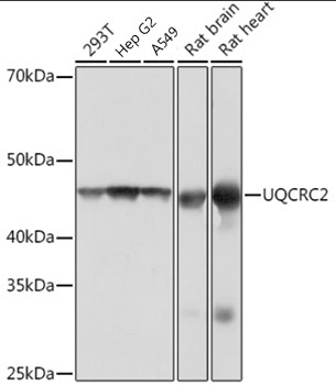
Western blot analysis of various lysates using UQCRC2 Rabbit mAb (CAB4366) at 1:1000 dilution. Secondary antibody: HRP-conjugated Goat anti-Rabbit IgG (H+L) (AS014) at 1:10000 dilution. Lysates/proteins: 25μg per lane. Blocking buffer: 3% nonfat dry milk in TBST. Detection: ECL Basic Kit (AbGn00020). Exposure time: 1s.
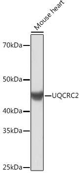
Western blot analysis of lysates from Mouse heart, using UQCRC2 Rabbit mAb (CAB4366) at 1:1000 dilution. Secondary antibody: HRP-conjugated Goat anti-Rabbit IgG (H+L) (AS014) at 1:10000 dilution. Lysates/proteins: 25μg per lane. Blocking buffer: 3% nonfat dry milk in TBST. Detection: ECL Basic Kit (AbGn00020). Exposure time: 3min.
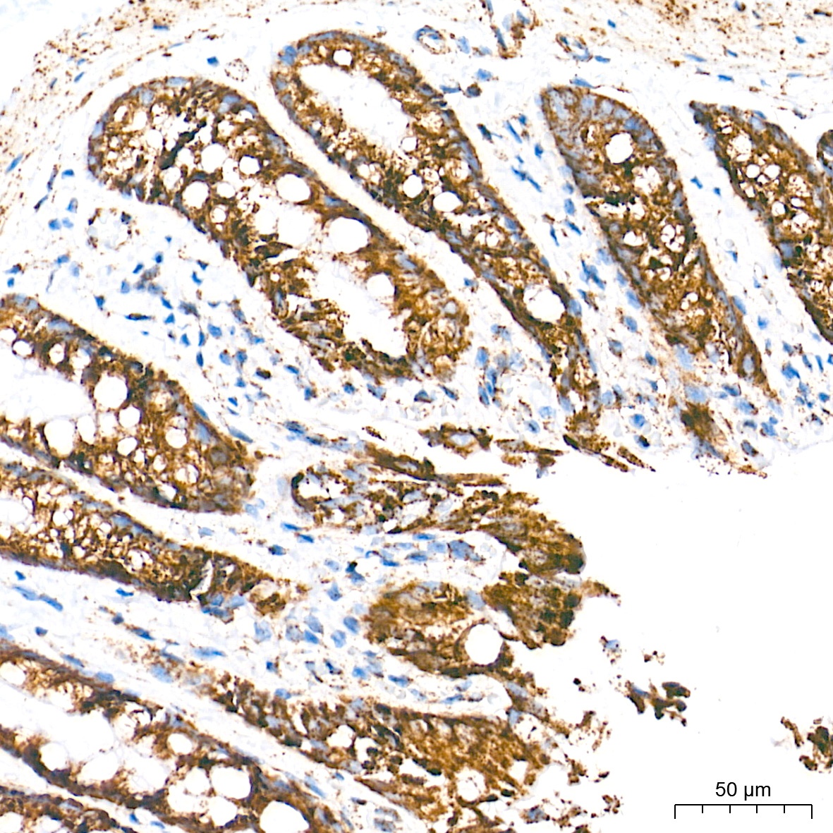
Immunohistochemistry analysis of paraffin-embedded Human colon carcinoma tissue using UQCRC2 Rabbit mAb (CAB4366) at a dilution of 1:500 (40x lens). High pressure antigen retrieval performed with 0.01M Tris-EDTA Buffer (pH 9.0) prior to IHC staining.
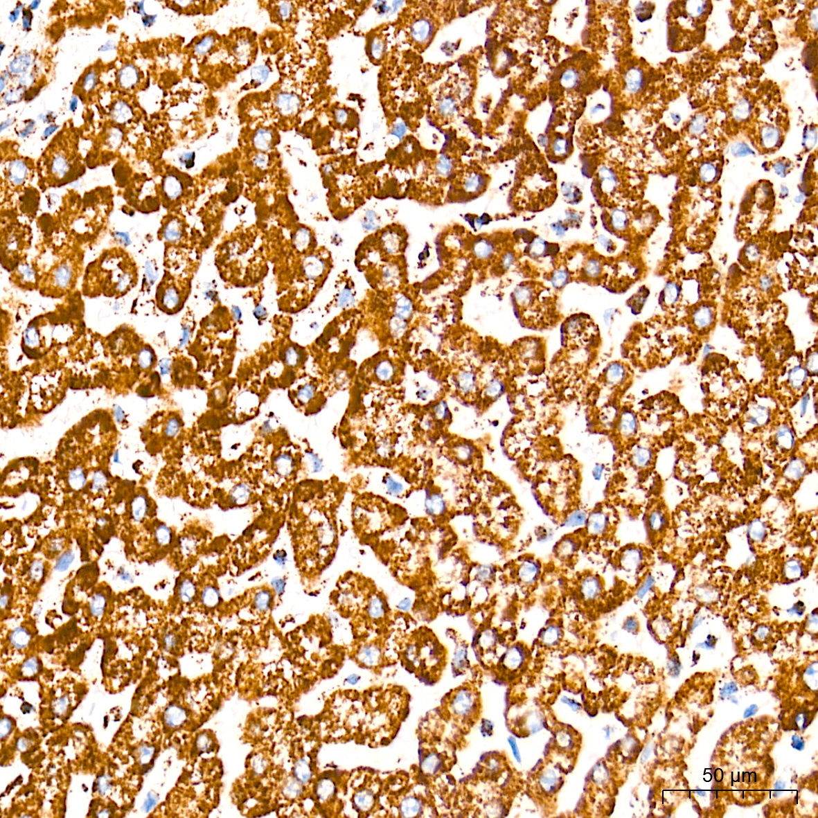
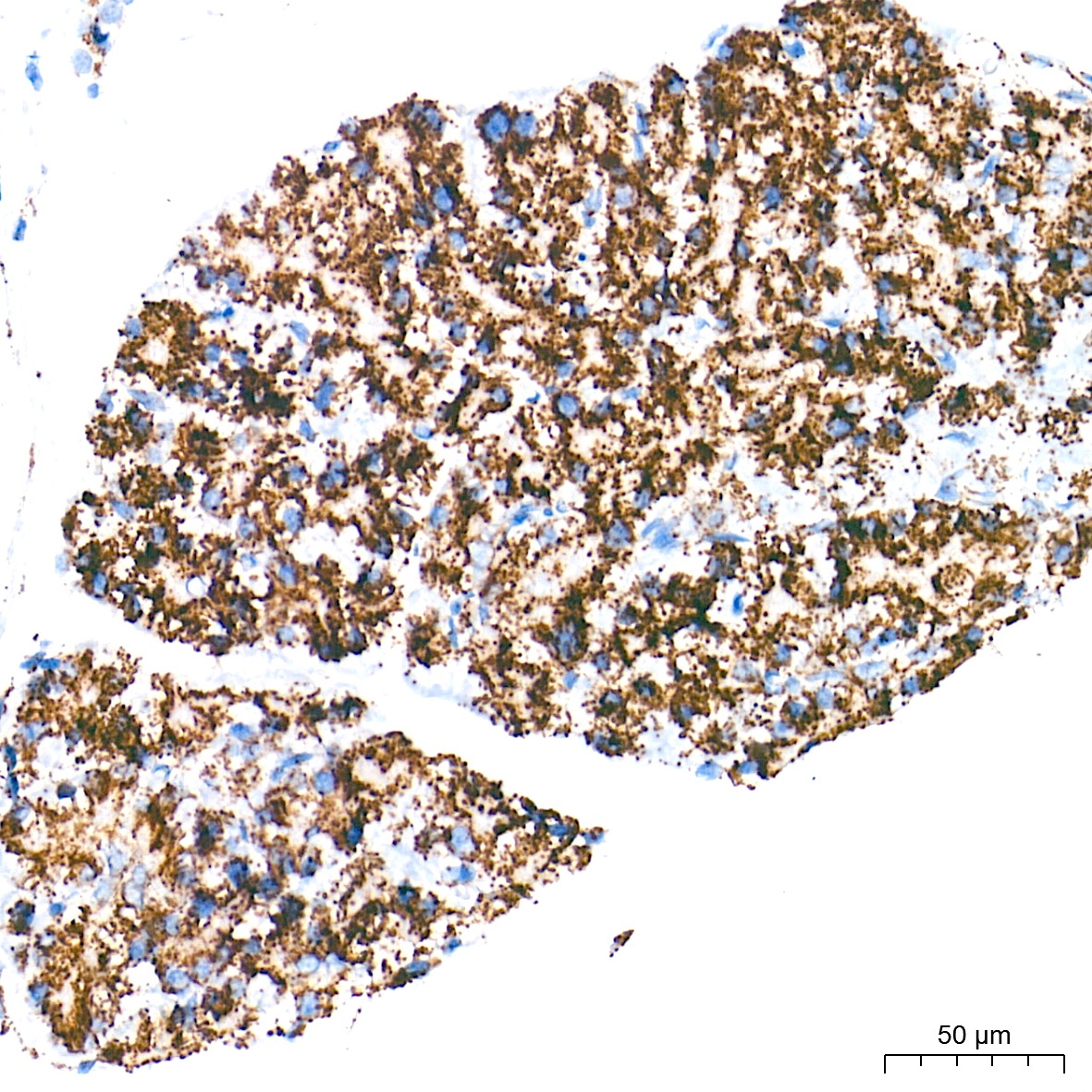
Immunohistochemistry analysis of paraffin-embedded Human liver tissue using UQCRC2 Rabbit mAb (CAB4366) at a dilution of 1:500 (40x lens). High pressure antigen retrieval performed with 0.01M Tris-EDTA Buffer (pH 9.0) prior to IHC staining.
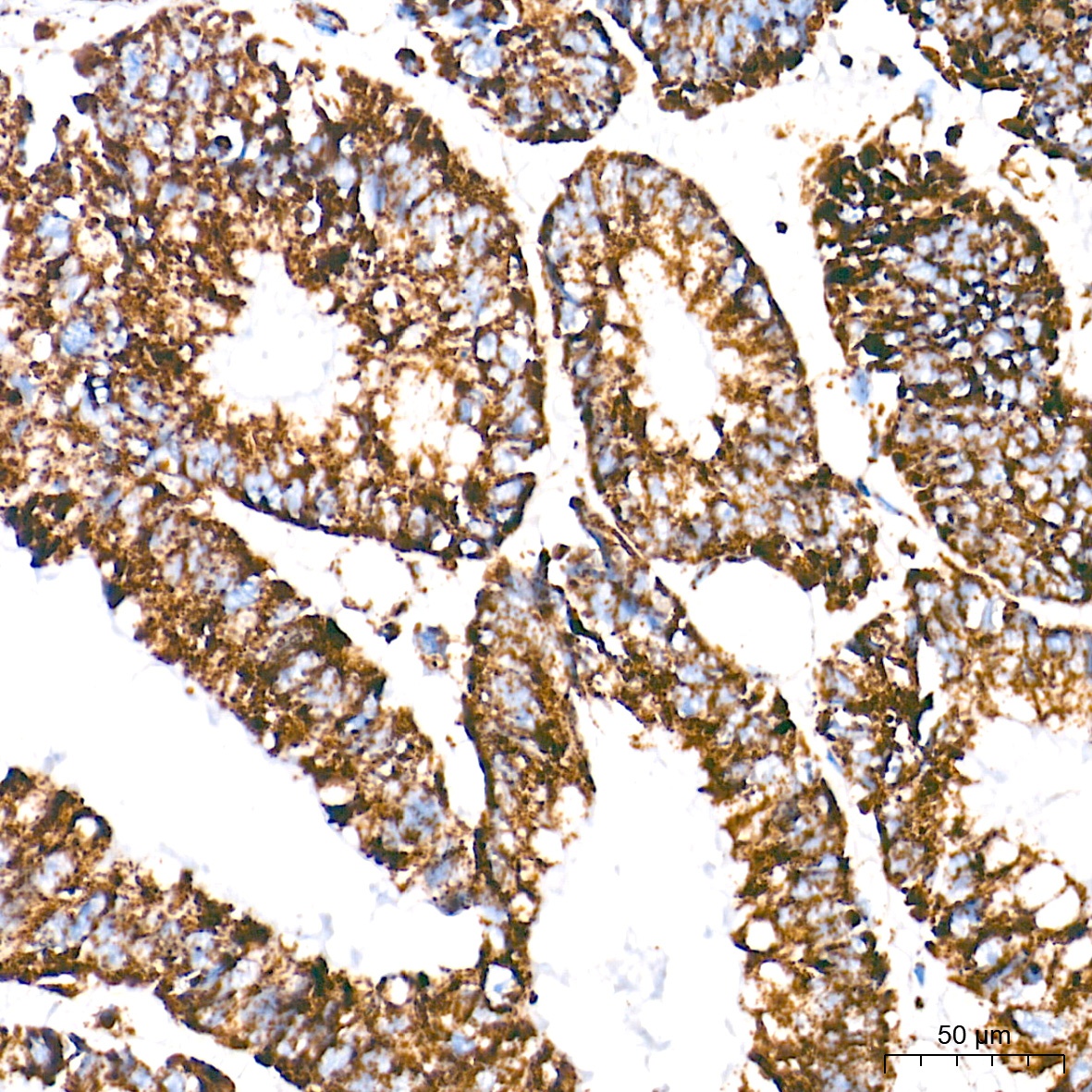
Immunohistochemistry analysis of paraffin-embedded Rat colon tissue using UQCRC2 Rabbit mAb (CAB4366) at a dilution of 1:500 (40x lens). High pressure antigen retrieval performed with 0.01M Tris-EDTA Buffer (pH 9.0) prior to IHC staining.
Immunohistochemistry analysis of paraffin-embedded Rat pancreas tissue using UQCRC2 Rabbit mAb (CAB4366) at a dilution of 1:500 (40x lens). High pressure antigen retrieval performed with 0.01M Tris-EDTA Buffer (pH 9.0) prior to IHC staining.