The VCP Antibody (CAB13368) is a high-quality antibody developed for reliable detection and analysis of target proteins. This cell surface molecule plays a crucial role in various cellular processes, including protein degradation and quality control. The antibody, produced in rabbits, exhibits high reactivity with human samples and has been validated for use in Western blot applications.By binding to the VCP protein, this antibody enables accurate detection and analysis in a variety of cell types. This makes it an essential tool for researchers studying cellular mechanisms, protein homeostasis, and diseases related to VCP dysfunction.
This antibody is validated for use in WB, IHC-P, IF/ICC, ELISA applications and has demonstrated reactivity against Human, Mouse, Rat samples.
Product Name:
VCP Antibody
SKU:
CAB13368
Size:
20μL, 100μL
Reactivity:
Human, Mouse, Rat
Conjugate:
Unconjugated
Immunogen:
Recombinant protein (or fragment).This information is considered to be commercially sensitive.
This gene encodes a member of the AAA ATPase family of proteins. The encoded protein plays a role in protein degradation, intracellular membrane fusion, DNA repair and replication, regulation of the cell cycle, and activation of the NF-kappa B pathway. This protein forms a homohexameric complex that interacts with a variety of cofactors and extracts ubiquitinated proteins from lipid membranes or protein complexes. Mutations in this gene cause IBMPFD (inclusion body myopathy with paget disease of bone and frontotemporal dementia), ALS (amyotrophic lateral sclerosis) and Charcot-Marie-Tooth disease in human patients.
Purification Method
Affinity purification
Gene ID
7415
RRID
AB_2760226
Buffer Information
Store at -20℃. Avoid freeze / thaw cycles. Buffer: PBS containing 50% glycerol, preserved with proclin300 or sodium azide, pH 7.3.
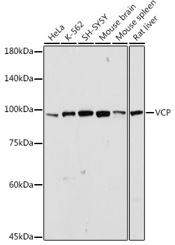
Western blot analysis of various lysates using VCP Rabbit pAb (CAB13368) at 1:1000 dilution. Secondary antibody: HRP-conjugated Goat anti-Rabbit IgG (H+L) (CABS014) at 1:10000 dilution. Lysates/proteins: 25μg per lane. Blocking buffer: 3% nonfat dry milk in TBST. Detection: ECL Basic Kit (AbGn00020). Exposure time: 30s.
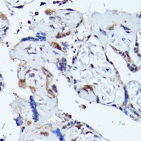
Immunohistochemistry analysis of paraffin-embedded Human placenta using VCP Rabbit pAb (CAB13368) at dilution of 1:100 (40x lens). High pressure antigen retrieval performed with 0.01M Citrate buffer (pH 6.0) prior to IHC staining.
Immunohistochemistry analysis of paraffin-embedded Rat kidney using VCP Rabbit pAb (CAB13368) at dilution of 1:100 (40x lens). High pressure antigen retrieval performed with 0.01M Citrate buffer (pH 6.0) prior to IHC staining.
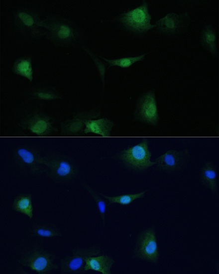
Immunofluorescence analysis of C6 cells using VCP Rabbit pAb (CAB13368) at dilution of 1:100. Secondary antibody: Cy3-conjugated Goat anti-Rabbit IgG (H+L) (CABS007) at 1:500 dilution. Blue: DAPI for nuclear staining.