The WIF1 Antibody (CAB5386) is a high-quality antibody developed for reliable detection and analysis of target proteins. The protein encoded by this gene functions to inhibit WNT proteins, which are extracellular signaling molecules that play a role in embryonic development. This protein contains a WNT inhibitory factor (WIF) domain and five epidermal growth factor (EGF)-like domains, and is thought to be involved in mesoderm segmentation. This gene functions as a tumor suppressor gene, and has been found to be epigenetically silenced in various cancers.
This antibody is validated for use in WB, IF/ICC, ELISA applications and has demonstrated reactivity against Human, Mouse, Rat samples.
Product Name:
WIF1 Antibody
SKU:
CAB5386
Size:
100μL, 20μL
Reactivity:
Human, Mouse, Rat
Conjugate:
Unconjugated
Immunogen:
Recombinant protein (or fragment).This information is considered to be commercially sensitive.
Tested Applications:
WBIF/ICCELISA
Recommended Dilution:
WB
1:100 - 1:500
IF/ICC
1:50 - 1:200
ELISA
Recommended starting concentration is 1 μg/mL. Please optimize the concentration based on your specific assay requirements.
Synonyms:
WIF-1, WIF1
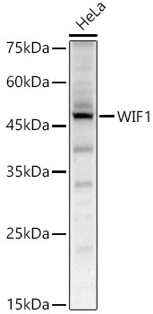
Positive Sample:
HeLa
Cellular Localization:
Secreted.
Calculated MW:
42kDa
Observed MW:
42kDa
The protein encoded by this gene functions to inhibit WNT proteins, which are extracellular signaling molecules that play a role in embryonic development. This protein contains a WNT inhibitory factor (WIF) domain and five epidermal growth factor (EGF)-like domains, and is thought to be involved in mesoderm segmentation. This gene functions as a tumor suppressor gene, and has been found to be epigenetically silenced in various cancers.
Purification Method
Affinity purification
Gene ID
11197
RRID
AB_2766195
Buffer Information
Store at -20℃. Avoid freeze / thaw cycles. Buffer: PBS containing 50% glycerol, preserved with proclin300 or sodium azide, pH 7.3.
Western blot analysis of lysates from HeLa cells, using WIF1 Rabbit pAb (CAB5386) at 1:500 dilution. Secondary antibody: HRP-conjugated Goat anti-Rabbit IgG (H+L) (AS014) at 1:10000 dilution. Lysates/proteins: 25μg per lane. Blocking buffer: 3% nonfat dry milk in TBST. Detection: ECL Enhanced Kit (AbGn00021). Exposure time: 60s.
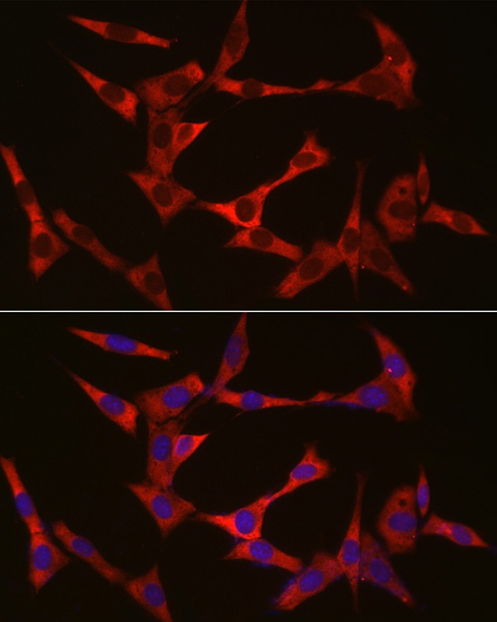
Immunofluorescence analysis of NIH/3T3 cells using WIF1 Rabbit pAb (CAB5386) at dilution of 1:50 (40x lens). Secondary antibody: Cy3-conjugated Goat anti-Rabbit IgG (H+L) (AS007) at 1:500 dilution. Blue: DAPI for nuclear staining.