The ZNF131 Antibody (CAB15331) is a high-quality antibody developed for reliable detection and analysis of target proteins. This antibody, generated in rabbits, is highly specific to human samples and has been validated for use in Western blot applications. By binding to the ZNF131 protein, this antibody allows for the detection and analysis of ZNF131 expression in various cell types, making it ideal for studies in molecular biology and genetics.ZNF131 is known to play a crucial role in transcriptional regulation and epigenetic modifications, making it a key player in gene expression and cell development.
This antibody is validated for use in WB, IF/ICC, ELISA applications and has demonstrated reactivity against Human, Mouse, Rat samples.
Product Name:
ZNF131 Antibody
SKU:
CAB15331
Size:
20μL, 100μL
Reactivity:
Human, Mouse, Rat
Conjugate:
Unconjugated
Immunogen:
Recombinant protein (or fragment).This information is considered to be commercially sensitive.
Recommended starting concentration is 1 μg/mL. Please optimize the concentration based on your specific assay requirements.
Synonyms:
ZBTB35, pHZ-10, ZNF131
Positive Sample:
293F
Cellular Localization:
Nucleus.
Calculated MW:
71kDa
Observed MW:
71kDa
Predicted to enable DNA-binding transcription activator activity, RNA polymerase II-specific; DNA-binding transcription repressor activity, RNA polymerase II-specific; and RNA polymerase II cis-regulatory region sequence-specific DNA binding activity. Predicted to be involved in positive regulation of transcription by RNA polymerase II. Located in intermediate filament cytoskeleton and nucleoplasm.
Purification Method
Affinity purification
Gene ID
7690
RRID
AB_2762233
Buffer Information
Store at -20℃. Avoid freeze / thaw cycles. Buffer: PBS containing 50% glycerol, preserved with proclin300 or sodium azide, pH 7.3.
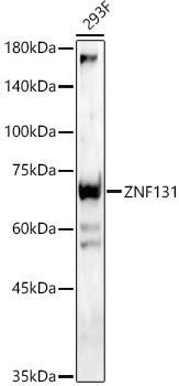
Western blot analysis of lysates from 293F cells, using ZNF131 Rabbit pAb (CAB15331) at 1:900 dilution. Secondary antibody: HRP-conjugated Goat anti-Rabbit IgG (H+L) (CABS014) at 1:10000 dilution. Lysates/proteins: 25μg per lane. Blocking buffer: 3% nonfat dry milk in TBST. Detection: ECL Enhanced Kit (AbGn00021). Exposure time: 30s.
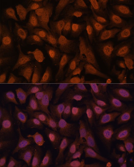
Immunofluorescence analysis of U-2 OS cells using ZNF131 Rabbit pAb (CAB15331) at dilution of 1:100. Secondary antibody: Cy3-conjugated Goat anti-Rabbit IgG (H+L) (CABS007) at 1:500 dilution. Blue: DAPI for nuclear staining.