The Histone H1.2 Monoclonal Antibody (CAB0646) is a high-quality antibody developed for reliable detection and analysis of target proteins. This polyclonal antibody, produced in rabbits, is highly specific to human histone H1.2 and has been validated for use in various applications, including Western blotting.Histone H1.2 is essential for maintaining chromatin structure and packaging DNA in the nucleus. Its role in gene expression regulation makes it a crucial player in cell differentiation, development, and disease progression.
This antibody is validated for use in WB, IF/ICC, ELISA applications and has demonstrated reactivity against Human, Other (Wide Range Predicted) samples.
Product Name:
Histone H1.2 Monoclonal Antibody
SKU:
CAB0646
Size:
20μL, 100μL
Reactivity:
Human, Other (Wide Range Predicted)
Clone Number:
ARC1836
Conjugate:
Unconjugated
Immunogen:
Synthetic peptide. This information is considered to be commercially sensitive.
Recommended starting concentration is 1 μg/mL. Please optimize the concentration based on your specific assay requirements.
Synonyms:
H1C, H1.2, H1F2, H1s-1, HIST1H1C, Histone H1.2
Positive Sample:
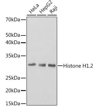
HeLa, Hep G2, Raji
Cellular Localization:
Nucleus.
Calculated MW:
21kDa
Observed MW:
32kDa
Histones are basic nuclear proteins responsible for nucleosome structure of the chromosomal fiber in eukaryotes. Two molecules of each of the four core histones (H2A, H2B, H3, and H4) form an octamer, around which approximately 146 bp of DNA is wrapped in repeating units, called nucleosomes. The linker histone, H1, interacts with linker DNA between nucleosomes and functions in the compaction of chromatin into higher order structures. This gene is intronless and encodes a replication-dependent histone that is a member of the histone H1 family. Transcripts from this gene lack polyA tails but instead contain a palindromic termination element. This gene is found in the large histone gene cluster on chromosome 6.
Purification Method
Affinity purification
Gene ID
3006
Buffer Information
Store at -20℃. Avoid freeze / thaw cycles. Buffer: PBS containing 50% glycerol and 0.05% BSA, preserved with proclin300 or sodium azide, pH 7.3.
Western blot analysis of various lysates using Histone H1.2 Rabbit mAb (CAB0646) at 1:3000 dilution. Secondary antibody: HRP-conjugated Goat anti-Rabbit IgG (H+L) (CABS014) at 1:10000 dilution. Lysates/proteins: 25μg per lane. Blocking buffer: 3% nonfat dry milk in TBST. Detection: ECL Basic Kit (AbGn00020). Exposure time: 1s.
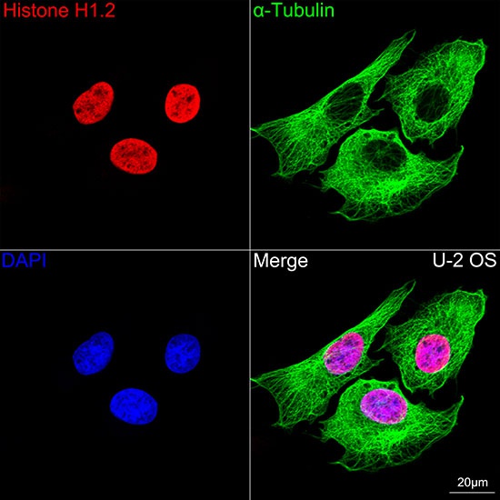
Confocal imaging of U-2 OS cells using Histone H1.2 Rabbit mAb (CAB0646,dilution 1:100)(Red). The cells were counterstained with α-Tubulin Mouse mAb (AC012,dilution 1:400) (Green). DAPI was used for nuclear staining (blue). Objective: 100x.


 ELISA Kit (AEFI00158)")
 ELISA Kit (AEFI00158)")

