The Acetyl-Histone H2B-K12 Antibody (CAB15619) is a high-quality antibody developed for reliable detection and analysis of target proteins. Histones are basic nuclear proteins that are responsible for the nucleosome structure of the chromosomal fiber in eukaryotes. Two molecules of each of the four core histones (H2A, H2B, H3, and H4) form an octamer, around which approximately 146 bp of DNA is wrapped in repeating units, called nucleosomes. The linker histone, H1, interacts with linker DNA between nucleosomes and functions in the compaction of chromatin into higher order structures. This gene encodes a replication-dependent histone that is a member of the histone H2B family, and generates two transcripts through the use of the conserved stem-loop termination motif, and the polyA addition motif. The protein has antibacterial and antifungal antimicrobial activity.
This antibody is validated for use in WB, IHC-P, IF/ICC, ChIP, ELISA applications and has demonstrated reactivity against Human, Mouse, Rat, Other (Wide Range Predicted) samples.
Product Name:
Acetyl-Histone H2B-K12 Antibody
SKU:
CAB15619
Size:
100μL, 20μL
Reactivity:
Human, Mouse, Rat, Other (Wide Range Predicted)
Conjugate:
Unconjugated
Immunogen:
Synthetic peptide. This information is considered to be commercially sensitive.
Tested Applications:
WBIHC-PIF/ICCChIPELISA
Recommended Dilution:
WB
1:500 - 1:1000
IHC-P
1:50 - 1:200
IF/ICC
1:50 - 1:200
ELISA
Recommended starting concentration is 1 μg/mL. Please optimize the concentration based on your specific assay requirements.
HeLa treated with TSA, C2C12 treated with TSA, C6 treated with TSA
Cellular Localization:
Chromosome, Nucleus.
Calculated MW:
14kDa
Observed MW:
14kDa
Histones are basic nuclear proteins that are responsible for the nucleosome structure of the chromosomal fiber in eukaryotes. Two molecules of each of the four core histones (H2A, H2B, H3, and H4) form an octamer, around which approximately 146 bp of DNA is wrapped in repeating units, called nucleosomes. The linker histone, H1, interacts with linker DNA between nucleosomes and functions in the compaction of chromatin into higher order structures. This gene encodes a replication-dependent histone that is a member of the histone H2B family, and generates two transcripts through the use of the conserved stem-loop termination motif, and the polyA addition motif. The protein has antibacterial and antifungal antimicrobial activity.
Purification Method
Affinity purification
Gene ID
3017 8349
RRID
AB_2763025
Buffer Information
Store at -20℃. Avoid freeze / thaw cycles. Buffer: PBS with 0.01% thimerosal,50% glycerol,pH7.3.
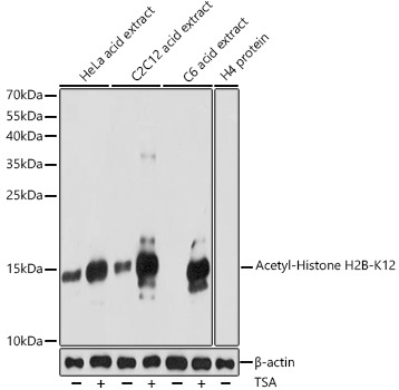
Western blot analysis of various lysates using Acetyl-Histone H2B-K12 Rabbit pAb (CAB15619) at 1:1000 dilution. HeLa cells and C2C12 cells and C6 cells were treated with TSA (1 uM) at 37℃ for 18 hours. Secondary antibody: HRP-conjugated Goat anti-Rabbit IgG (H+L) (AS014) at 1:10000 dilution. Lysates/proteins: 25μg per lane. Blocking buffer: 3% nonfat dry milk in TBST. Detection: ECL Basic Kit (AbGn00020). Exposure time: 90s.
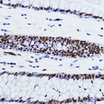
Immunohistochemistry analysis of paraffin-embedded Human colon using Acetyl-Histone H2B-K12 Rabbit pAb (CAB15619) at dilution of 1:200 (40x lens). Microwave antigen retrieval performed with 0.01M PBS Buffer (pH 7.2) prior to IHC staining.
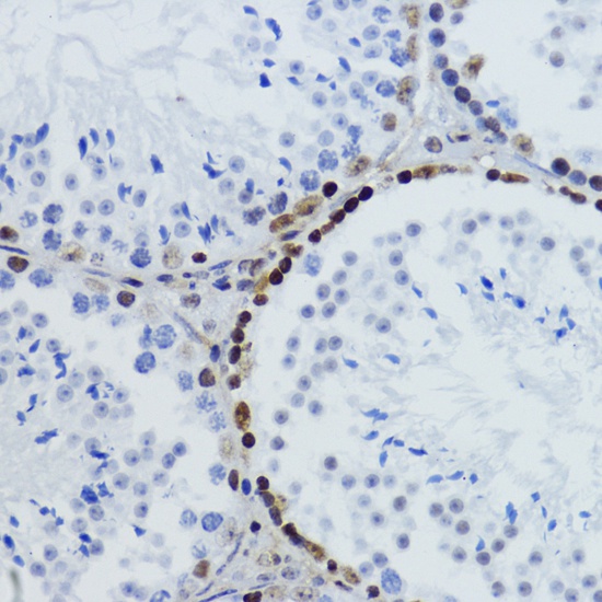
Immunohistochemistry analysis of paraffin-embedded Mouse testis using Acetyl-Histone H2B-K12 Rabbit pAb (CAB15619) at dilution of 1:200 (40x lens). Microwave antigen retrieval performed with 0.01M PBS Buffer (pH 7.2) prior to IHC staining.
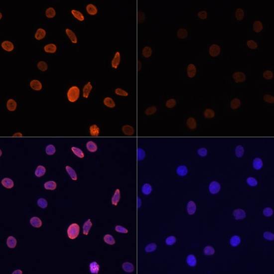
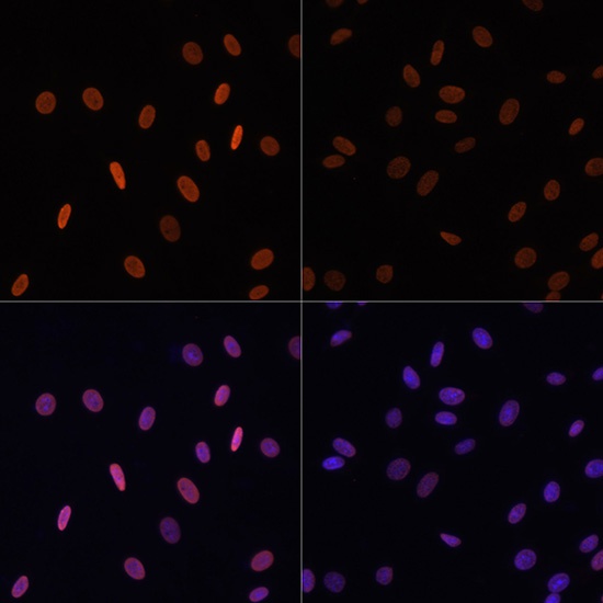
Immunofluorescence analysis of C6 cells using Acetyl-Histone H2B-K12 Rabbit pAb (CAB15619) at dilution of 1:100.C6 cells were treated with TSA (1 uM) at 37℃ for 18 hours (top left). Blue: DAPI for nuclear staining.
Immunofluorescence analysis of NIH/3T3 cells using Acetyl-Histone H2B-K12 Rabbit pAb (CAB15619) at dilution of 1:100.NIH/3T3 cells were treated with TSA (1 uM) at 37℃ for 18 hours (top left). Blue: DAPI for nuclear staining.
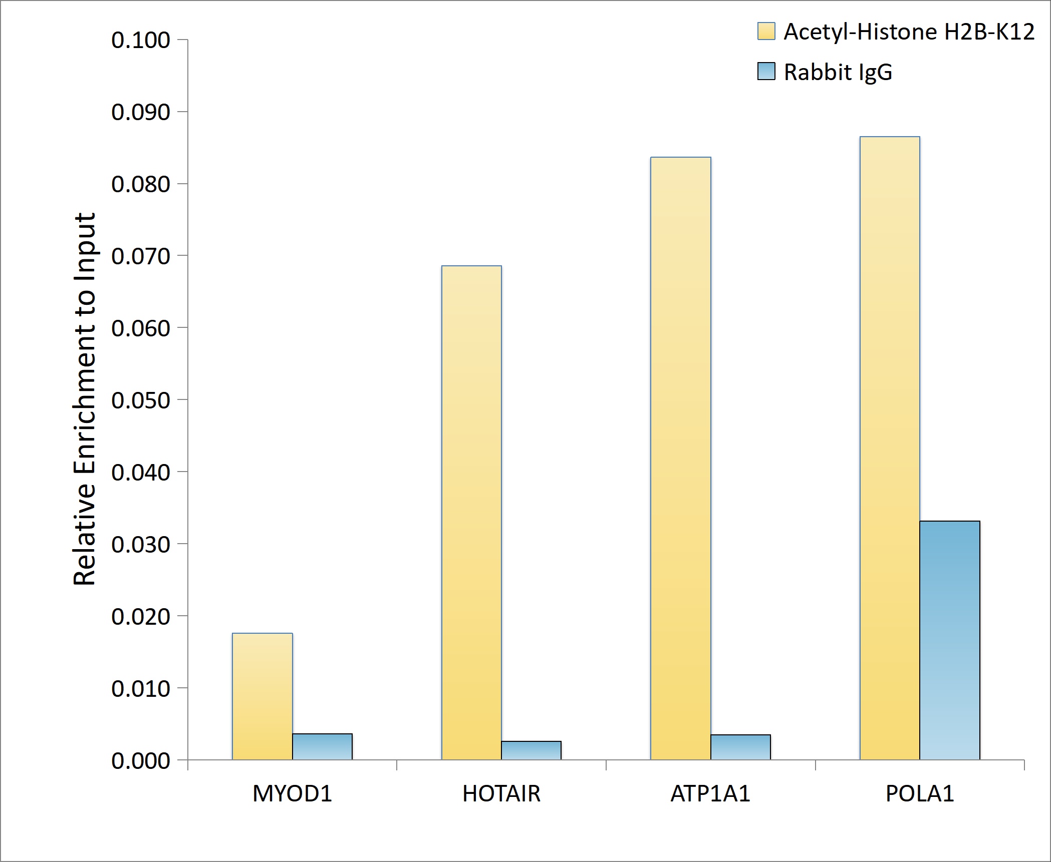
Chromatin immunoprecipitation analysis of extracts of HeLa cells, using Acetyl-Histone H2B-K12 antibody (CAB15619) and rabbit IgG.The amount of immunoprecipitated DNA was checked by quantitative PCR. Histogram was constructed by the ratios of the immunoprecipitated DNA to the input.