The AKAP8 Antibody (CAB3049) is a high-quality antibody developed for reliable detection and analysis of target proteins. This antibody is raised in rabbits and is highly specific to AKAP8 in human samples, making it suitable for applications such as Western blotting.AKAP8, also known as A-kinase anchor protein 8, is involved in various cellular processes, including cell growth, differentiation, and apoptosis. By targeting AKAP8 with this antibody, researchers can study its function in different cell types and gain insights into its potential role in disease mechanisms.
This antibody is validated for use in WB, IF/ICC, ELISA applications and has demonstrated reactivity against Human, Mouse samples.
Product Name:
AKAP8 Antibody
SKU:
CAB3049
Size:
20μL, 100μL
Reactivity:
Human, Mouse
Conjugate:
Unconjugated
Immunogen:
Recombinant protein (or fragment).This information is considered to be commercially sensitive.
Recommended starting concentration is 1 μg/mL. Please optimize the concentration based on your specific assay requirements.
Synonyms:
AKAP-8, AKAP95, AKAP 95, AKAP-95, AKAP8
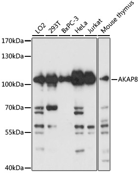
Positive Sample:
LO2, 293T, BxPC-3, HeLa, Jurkat, mouse thymus
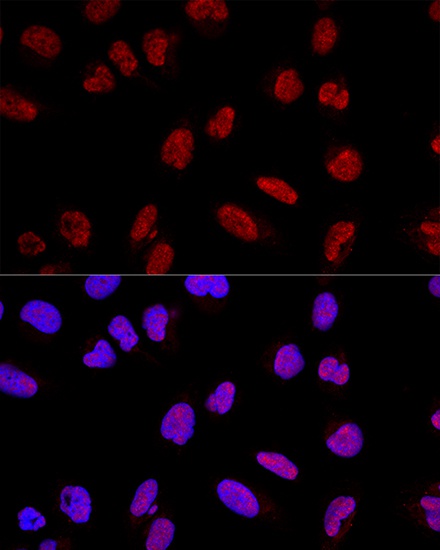
Cellular Localization:
Nucleus Matrix.
Calculated MW:
76kDa
Observed MW:
110kDa
This gene encodes a member of the A-kinase anchor protein family. A-kinase anchor proteins are scaffold proteins that contain a binding domain for the RI/RII subunit of protein kinase A (PKA) and recruit PKA and other signaling molecules to specific subcellular locations. This gene encodes a nuclear A-kinase anchor protein that binds to the RII alpha subunit of PKA and may play a role in chromosome condensation during mitosis by targeting PKA and the condensin complex to chromatin. A pseudogene of this gene is located on the short arm of chromosome 9.
Purification Method
Affinity purification
Gene ID
10270
RRID
AB_2764853
Buffer Information
Store at -20℃. Avoid freeze / thaw cycles. Buffer: PBS with 0.01% thimerosal,50% glycerol,pH7.3.
Western blot analysis of various lysates using AKAP8 Rabbit pAb (CAB3049) at 1:3000 dilution. Secondary antibody: HRP-conjugated Goat anti-Rabbit IgG (H+L) (CABS014) at 1:10000 dilution. Lysates/proteins: 25μg per lane. Blocking buffer: 3% nonfat dry milk in TBST. Detection: ECL Basic Kit (AbGn00020). Exposure time: 30s.
Confocal immunofluorescence analysis of U2OS cells using AKAP8 Rabbit pAb (CAB3049) at dilution of 1:200. Blue: DAPI for nuclear staining.