The ALDH1A1 Monoclonal Antibody (CAB0157) is a high-quality antibody developed for reliable detection and analysis of target proteins. This antibody, produced using rabbit monoclonal technology, exhibits high specificity and sensitivity for detecting ALDH1A1 in human samples, making it ideal for use in Western blot and immunohistochemistry applications.ALDH1A1, a member of the aldehyde dehydrogenase family, plays a key role in retinoic acid biosynthesis and is known to be a marker for cancer stem cells in various types of tumors.
This antibody is validated for use in WB, IF/ICC, IP, ELISA applications and has demonstrated reactivity against Human, Mouse, Rat samples.
Product Name:
ALDH1A1 Monoclonal Antibody
SKU:
CAB0157
Size:
20μL, 100μL
Reactivity:
Human, Mouse, Rat
Clone Number:
ARC52440
Conjugate:
Unconjugated
Immunogen:
A synthetic peptide corresponding to a sequence within amino acids 250-350 of human ALDH1A1 (NP_000680.2).
A549, Hep G2, Mouse lung, Mouse liver, Mouse testis, Rat lung, Rat kidney, Rat testis
Cellular Localization:
Cytoplasm.
Calculated MW:
55kDa
Observed MW:
55kDa
The protein encoded by this gene belongs to the aldehyde dehydrogenase family. Aldehyde dehydrogenase is the next enzyme after alcohol dehydrogenase in the major pathway of alcohol metabolism. There are two major aldehyde dehydrogenase isozymes in the liver, cytosolic and mitochondrial, which are encoded by distinct genes, and can be distinguished by their electrophoretic mobility, kinetic properties, and subcellular localization. This gene encodes the cytosolic isozyme. Studies in mice show that through its role in retinol metabolism, this gene may also be involved in the regulation of the metabolic responses to high-fat diet.
Purification Method
Affinity purification
Gene ID
216
RRID
AB_2861455
Buffer Information
Store at -20℃. Avoid freeze / thaw cycles. Buffer: PBS with 0.09% Sodium azide,0.05% BSA,50% glycerol,pH7.3.
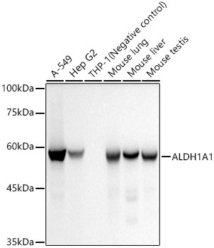
Western blot analysis of various lysates, using ALDH1A1 Rabbit mAb (CAB0157) at 1:20000 dilution. Secondary antibody: HRP-conjugated Goat anti-Rabbit IgG (H+L) (CABS014) at 1:10000 dilution. Lysates/proteins: 25μg per lane. Blocking buffer: 3% nonfat dry milk in TBST. Detection: ECL Basic Kit (AbGn00020). Negative control (NC): THP-1 Exposure time: 10s.
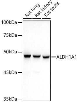
Western blot analysis of various lysates, using ALDH1A1 Rabbit mAb (CAB0157) at 1:20000 dilution. Secondary antibody: HRP-conjugated Goat anti-Rabbit IgG (H+L) (CABS014) at 1:10000 dilution. Lysates/proteins: 25μg per lane. Blocking buffer: 3% nonfat dry milk in TBST. Detection: ECL Basic Kit (AbGn00020). Exposure time: 90s.
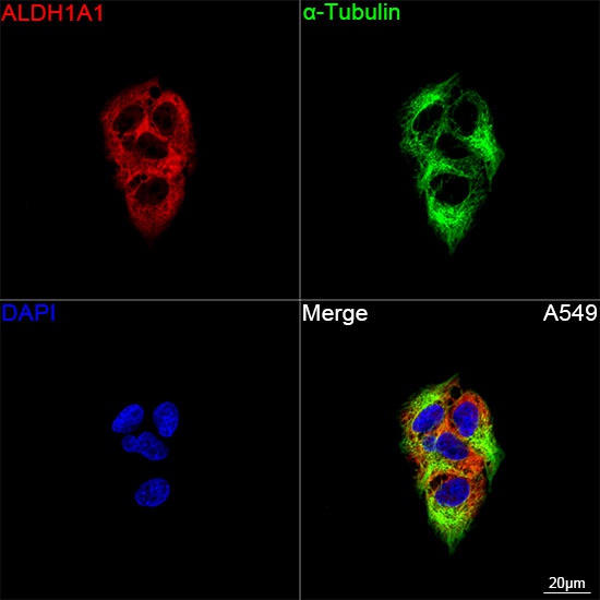
Confocal imaging of A549 cells using ALDH1A1 Rabbit mAb (CAB0157, dilution 1:8000) followed by a further incubation with Cy3 Goat Anti-Rabbit IgG (H+L) (CABS007, dilution 1:500) (Red). The cells were counterstained with α-Tubulin Mouse mAb (AC012, dilution 1:400) followed by incubation with ABflo® 488-conjugated Goat Anti-Mouse IgG (H+L) Ab (CABS076, dilution 1:500) (Green). DAPI was used for nuclear staining (Blue). Objective: 100x.
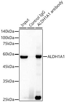
Immunoprecipitation analysis of 300 μg extracts of A-549 cells using 3 μg ALDH1A1 Rabbit mAb antibody (CAB0157). Western blot was performed from the immunoprecipitate using ALDH1A1 Rabbit mAb (CAB0157) at a dilition of 1:20000.