The ALDH1A1 Antibody (CAB1802) is a high-quality antibody developed for reliable detection and analysis of target proteins. This antibody, produced in rabbits, is highly specific to human samples and has been validated for use in Western blot applications. By binding to the ALDH1A1 protein, this antibody enables precise detection and analysis in a variety of cell types, making it an essential tool for investigations in stem cell biology, cancer research, and drug development.ALDH1A1, also known as aldehyde dehydrogenase 1 family member A1, plays a crucial role in maintaining cellular homeostasis and differentiation processes.
This antibody is validated for use in WB, IHC-P, IF/ICC, ELISA applications and has demonstrated reactivity against Human, Mouse, Rat samples.
Product Name:
ALDH1A1 Antibody
SKU:
CAB1802
Size:
20μL, 100μL
Reactivity:
Human, Mouse, Rat
Conjugate:
Unconjugated
Immunogen:
Synthetic peptide. This information is considered to be commercially sensitive.
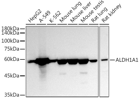
HepG2, A-549, K-562, Mouse lung, Mouse liver, Mouse testis, Rat lung, Rat kidney
Cellular Localization:
Cytoplasm.
Calculated MW:
55kDa
Observed MW:
55kDa
The protein encoded by this gene belongs to the aldehyde dehydrogenase family. Aldehyde dehydrogenase is the next enzyme after alcohol dehydrogenase in the major pathway of alcohol metabolism. There are two major aldehyde dehydrogenase isozymes in the liver, cytosolic and mitochondrial, which are encoded by distinct genes, and can be distinguished by their electrophoretic mobility, kinetic properties, and subcellular localization. This gene encodes the cytosolic isozyme. Studies in mice show that through its role in retinol metabolism, this gene may also be involved in the regulation of the metabolic responses to high-fat diet.
Purification Method
Affinity purification
Gene ID
216
RRID
AB_2763841
Buffer Information
Store at -20℃. Avoid freeze / thaw cycles. Buffer: PBS containing 50% glycerol, preserved with proclin300 or sodium azide, pH 7.3.
Western blot analysis of various lysates using ALDH1A1 Rabbit pAb (CAB1802) at 1:1000 dilution. Secondary antibody: HRP-conjugated Goat anti-Rabbit IgG (H+L) (CABS014) at 1:10000 dilution. Lysates/proteins: 25μg per lane. Blocking buffer: 3% nonfat dry milk in TBST. Detection: ECL Basic Kit (AbGn00020). Exposure time: 30s.
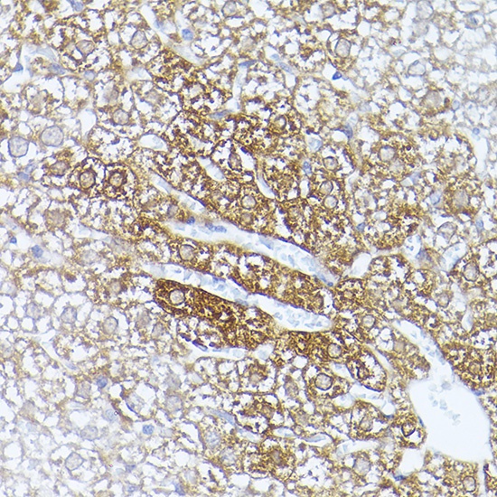
Immunohistochemistry analysis of paraffin-embedded Mouse liver using ALDH1A1 Rabbit pAb (CAB1802) at dilution of 1:20 (40x lens). High pressure antigen retrieval performed with 0.01M Citrate buffer (pH 6.0) prior to IHC staining.
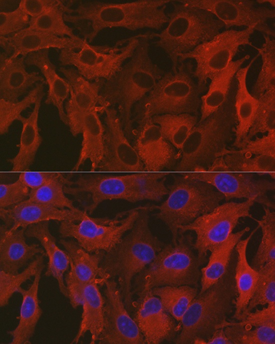
Immunofluorescence analysis of A-549 cells using ALDH1A1 Rabbit pAb (CAB1802) at dilution of 1:100 (40x lens). Secondary antibody: Cy3-conjugated Goat anti-Rabbit IgG (H+L) (CABS007) at 1:500 dilution. Blue: DAPI for nuclear staining.
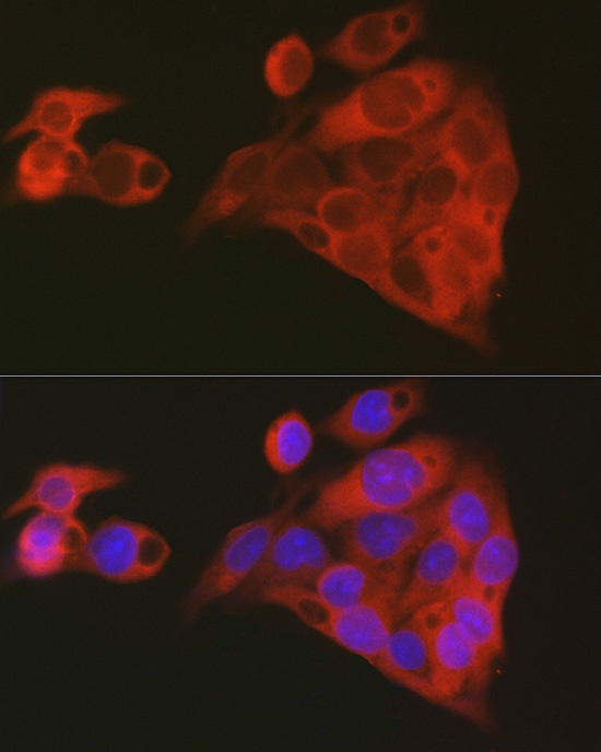
Immunofluorescence analysis of HepG2 cells using ALDH1A1 Rabbit pAb (CAB1802) at dilution of 1:100 (40x lens). Secondary antibody: Cy3-conjugated Goat anti-Rabbit IgG (H+L) (CABS007) at 1:500 dilution. Blue: DAPI for nuclear staining.