The Cardiac Troponin C Monoclonal Antibody (CAB3816) is a high-quality antibody developed for reliable detection and analysis of target proteins. This polyclonal antibody, generated in rabbits, is highly specific and reactive with human samples, making it an excellent choice for Western blot applications.Cardiac troponin C is a key component of the troponin complex, which plays a critical role in regulating muscle contraction in the heart. Elevated levels of cardiac troponin C in the blood are a well-established marker of cardiac injury, making it a valuable target for diagnostic and research purposes in cardiovascular disease.
This antibody is validated for use in WB, ELISA applications and has demonstrated reactivity against Mouse, Rat samples.
Product Name:
Cardiac Troponin C Monoclonal Antibody
SKU:
CAB3816
Size:
20μL, 100μL
Reactivity:
Mouse, Rat
Clone Number:
ARC0840
Conjugate:
Unconjugated
Immunogen:
Synthetic peptide. This information is considered to be commercially sensitive.
Recommended starting concentration is 1 μg/mL. Please optimize the concentration based on your specific assay requirements.
Synonyms:
TNC, TN-C, TNNC, CMD1Z, CMH13, TNNC1
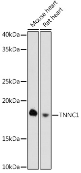
Positive Sample:
Mouse heart, Rat heart
Cellular Localization:
Cytosol.
Calculated MW:
18kDa
Observed MW:
18kDa
Troponin is a central regulatory protein of striated muscle contraction, and together with tropomyosin, is located on the actin filament. Troponin consists of 3 subunits: TnI, which is the inhibitor of actomyosin ATPase; TnT, which contains the binding site for tropomyosin; and TnC, the protein encoded by this gene. The binding of calcium to TnC abolishes the inhibitory action of TnI, thus allowing the interaction of actin with myosin, the hydrolysis of ATP, and the generation of tension. Mutations in this gene are associated with cardiomyopathy dilated type 1Z.
Purification Method
Affinity purification
Gene ID
7134
Buffer Information
Store at -20℃. Avoid freeze / thaw cycles. Buffer: PBS containing 50% glycerol and 0.05% BSA, preserved with proclin300 or sodium azide, pH 7.3.
Western blot analysis of various lysates using TNNC1 Rabbit mAb (CAB3816) at 1:1000 dilution. Secondary antibody: HRP-conjugated Goat anti-Rabbit IgG (H+L) (CABS014) at 1:10000 dilution. Lysates/proteins: 25μg per lane. Blocking buffer: 3% nonfat dry milk in TBST. Detection: ECL Basic Kit (AbGn00020). Exposure time: 180s.