The CHM Monoclonal Antibody (CAB9378) is a high-quality antibody developed for reliable detection and analysis of target proteins. This antibody, generated in rabbits, exhibits high reactivity with human samples and has been validated for use in Western blot applications. By binding specifically to the CHM protein, researchers can easily detect and analyze its expression in various cell types.CHM, also known as CHM protein or REP1, plays a crucial role in regulating membrane trafficking pathways and cellular functions. Dysregulation of CHM has been linked to cancer progression, making it a potential therapeutic target for cancer treatment.
This antibody is validated for use in WB, ELISA applications and has demonstrated reactivity against Human samples.
Product Name:
CHM Monoclonal Antibody
SKU:
CAB9378
Size:
20μL, 100μL
Reactivity:
Human
Clone Number:
ARC2736
Conjugate:
Unconjugated
Immunogen:
Synthetic peptide. This information is considered to be commercially sensitive.
Recommended starting concentration is 1 μg/mL. Please optimize the concentration based on your specific assay requirements.
Synonyms:
TCD, GGTA, REP-1, DXS540, HSD-32, CHM
Positive Sample:
293T
Cellular Localization:
Cytoplasm, Cytosol.
Calculated MW:
73kDa
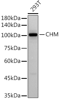
Observed MW:
101kDa
This gene encodes component A of the RAB geranylgeranyl transferase holoenzyme. In the dimeric holoenzyme, this subunit binds unprenylated Rab GTPases and then presents them to the catalytic Rab GGTase subunit for the geranylgeranyl transfer reaction. Rab GTPases need to be geranylgeranyled on either one or two cysteine residues in their C-terminus to localize to the correct intracellular membrane. Mutations in this gene are a cause of choroideremia; also known as tapetochoroidal dystrophy (TCD). This X-linked disease is characterized by progressive dystrophy of the choroid, retinal pigment epithelium and retina. Alternatively spliced transcript variants have been found for this gene.
Purification Method
Affinity purification
Gene ID
1121
Buffer Information
Store at -20℃. Avoid freeze / thaw cycles. Buffer: PBS containing 50% glycerol and 0.05% BSA, preserved with proclin300 or sodium azide, pH 7.3.
Western blot analysis of lysates from 293T cells, using (CAB9378) at 1:500 dilution. Secondary antibody: HRP-conjugated Goat anti-Rabbit IgG (H+L) (CABS014) at 1:10000 dilution. Lysates/proteins: 25μg per lane. Blocking buffer: 3% nonfat dry milk in TBST. Detection: ECL Basic Kit (AbGn00020). Exposure time: 60s.
 at 1:10000 dilution. Lysates/proteins: 25ug per lane. Blocking buffer: 3% nonfat dry milk in TBST. Detection: ECL Basic Kit. Exposure time: 60s.")
 at 1:10000 dilution. Lysates/proteins: 25ug per lane. Blocking buffer: 3% nonfat dry milk in TBST. Detection: ECL Basic Kit. Exposure time: 60s.")
![Anti-CHM [R36-7A-8] Monoclonal Antibody (AGMB03422)](https://cdn11.bigcommerce.com/s-h68l9z2lnx/images/stencil/590x590/products/274711/680419/anti-chm-r36-7a-8-monoclonal-antibody-agmb03422__29325.1773042022.jpg?c=2 "Anti-CHM [R36-7A-8] Monoclonal Antibody (AGMB03422)")


