The EAAT1/SLC1A3 Monoclonal Antibody (CAB9712) is a high-quality antibody developed for reliable detection and analysis of target proteins. This polyclonal antibody, produced in rabbits, is highly specific to human samples and has been validated for use in Western blot applications.EAAT1 is a crucial protein involved in the regulation of glutamate levels in the central nervous system, and dysregulation of EAAT1 has been linked to various neurological disorders such as epilepsy, Alzheimer's disease, and amyotrophic lateral sclerosis (ALS).
This antibody is validated for use in WB, IHC-P, ELISA, IF-P applications and has demonstrated reactivity against Mouse, Rat samples.
Product Name:
EAAT1/SLC1A3 Monoclonal Antibody
SKU:
CAB9712
Size:
20μL, 100μL
Reactivity:
Mouse, Rat
Clone Number:
ARC1714
Conjugate:
Unconjugated
Immunogen:
Synthetic peptide. This information is considered to be commercially sensitive.
Recommended starting concentration is 1 μg/mL. Please optimize the concentration based on your specific assay requirements.
Synonyms:
EA6, EAAT1, GLAST, GLAST1, EAAT1/SLC1A3
Positive Sample:
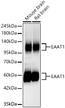
Mouse brain, Rat brain
Cellular Localization:
Membrane, Multi-Pass Membrane Protein.
Calculated MW:
60kDa
Observed MW:
50-55kDa/90-130kDa
This gene encodes a member of a member of a high affinity glutamate transporter family. This gene functions in the termination of excitatory neurotransmission in central nervous system. Mutations are associated with episodic ataxia, Type 6. Alternative splicing results in multiple transcript variants.
Purification Method
Affinity purification
Gene ID
6507
RRID
AB_2863763
Buffer Information
Store at -20℃. Avoid freeze / thaw cycles. Buffer: PBS containing 50% glycerol and 0.05% BSA, preserved with proclin300 or sodium azide, pH 7.3.
Western blot analysis of various lysates using EAAT1/SLC1A3 Rabbit mAb (CAB9712) at 1:1000 dilution incubated overnight at 4℃. Secondary antibody: HRP-conjugated Goat anti-Rabbit IgG (H+L) (CABS014) at 1:10000 dilution. Lysates/proteins: 25 μg per lane. Blocking buffer: 3% nonfat dry milk in TBST. Detection: ECL Basic Kit (AbGn00020). Exposure time: 45s.
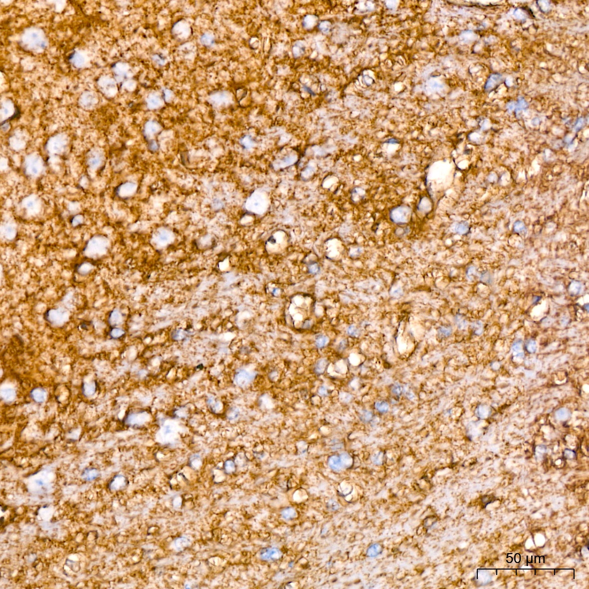
Immunohistochemistry analysis of paraffin-embedded Mouse brain tissue using EAAT1/SLC1A3 Rabbit mAb (CAB9712) at a dilution of 1:200 (40x lens). High pressure antigen retrieval performed with 0.01M Citrate buffer (pH 6.0) prior to IHC staining.
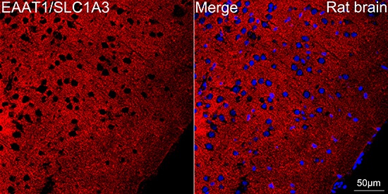
Confocal imaging of paraffin-embedded Rat brain tissue using EAAT1/SLC1A3 Rabbit mAb (CAB9712, dilution 1:200) followed by a further incubation with Cy3 Goat Anti-Rabbit IgG (H+L) (CABS007, dilution 1:500) (Red). DAPI was used for nuclear staining (Blue). Objective: 40x. Perform microwave antigen retrieval with 0.01 M citrate buffer (pH 6.0) prior to IF staining.
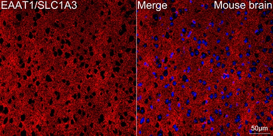
Confocal imaging of paraffin-embedded Mouse brain tissue using EAAT1/SLC1A3 Rabbit mAb (CAB9712, dilution 1:200) followed by a further incubation with Cy3 Goat Anti-Rabbit IgG (H+L) (CABS007, dilution 1:500) (Red). DAPI was used for nuclear staining (Blue). Objective: 40x. Perform microwave antigen retrieval with 0.01 M citrate buffer (pH 6.0) prior to IF staining.


")
")
![Anti-EAAT1 [R05-5D9] Monoclonal Antibody (AGMB01410)](https://cdn11.bigcommerce.com/s-h68l9z2lnx/images/stencil/590x590/products/272699/679209/anti-eaat1-r05-5d9-monoclonal-antibody-agmb01410__73920.1773038187.jpg?c=2 "Anti-EAAT1 [R05-5D9] Monoclonal Antibody (AGMB01410)")


