The PDIA4 Antibody (CAB4326) is a high-quality antibody developed for reliable detection and analysis of target proteins. This antibody, produced in rabbits, exhibits high specificity and sensitivity towards human samples, making it ideal for Western blot applications.PDIA4, also known as protein disulfide-isomerase A4, is essential for proper protein folding and assembly, ensuring the correct structure and function of proteins within the cell. Dysregulation of PDIA4 has been implicated in various diseases, including cancer, neurodegenerative disorders, and metabolic conditions, highlighting its potential as a therapeutic target.
This antibody is validated for use in WB, IHC-P, IF/ICC, ELISA applications and has demonstrated reactivity against Human, Mouse, Rat samples.
Product Name:
PDIA4 Antibody
SKU:
CAB4326
Size:
20μL, 100μL
Reactivity:
Human, Mouse, Rat
Conjugate:
Unconjugated
Immunogen:
Synthetic peptide. This information is considered to be commercially sensitive.
Recommended starting concentration is 1 μg/mL. Please optimize the concentration based on your specific assay requirements.
Synonyms:
ERP70, ERP72, ERp-72, PDIA4
Positive Sample:
HepG2, MCF7, Mouse liver, Rat lung, Rat liver
Cellular Localization:
Endoplasmic Reticulum Lumen, Melanosome.
Calculated MW:
73kDa
Observed MW:
72kDa
This gene encodes a member of the disulfide isomerase (PDI) family of endoplasmic reticulum (ER) proteins that catalyze protein folding and thiol-disulfide interchange reactions. The encoded protein has an N-terminal ER-signal sequence, three catalytically active thioredoxin (TRX) domains, two TRX-like domains and a C-terminal ER-retention sequence. This protein, when bound to cyclophilin B, enhances the rate of immunoglobulin G intermolecular disulfide bonding and antibody assembly.
Purification Method
Affinity purification
Gene ID
9601
RRID
AB_2765625
Buffer Information
Store at -20℃. Avoid freeze / thaw cycles. Buffer: PBS containing 50% glycerol, preserved with proclin300 or sodium azide, pH 7.3.
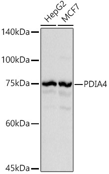
Western blot analysis of various lysates using PDIA4 Rabbit pAb (CAB4326) at 1:1000 dilution. Secondary antibody: HRP-conjugated Goat anti-Rabbit IgG (H+L) (CABS014) at 1:10000 dilution. Lysates/proteins: 25μg per lane. Blocking buffer: 3% nonfat dry milk in TBST. Detection: ECL Basic Kit (AbGn00020). Exposure time: 0.5s.
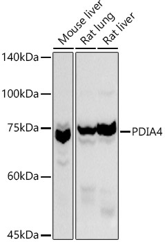
Western blot analysis of various lysates using PDIA4 Rabbit pAb (CAB4326) at 1:1000 dilution. Secondary antibody: HRP-conjugated Goat anti-Rabbit IgG (H+L) (CABS014) at 1:10000 dilution. Lysates/proteins: 25μg per lane. Blocking buffer: 3% nonfat dry milk in TBST. Detection: ECL Basic Kit (AbGn00020). Exposure time: 10s.
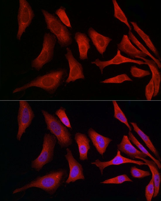
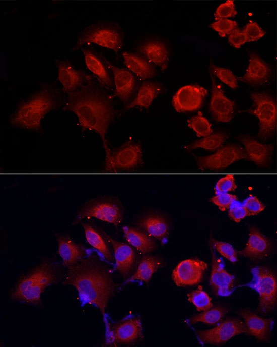
Immunofluorescence analysis of HeLa cells using PDIA4 Rabbit pAb (CAB4326) at dilution of 1:50 (40x lens). Secondary antibody: Cy3-conjugated Goat anti-Rabbit IgG (H+L) (CABS007) at 1:500 dilution. Blue: DAPI for nuclear staining.
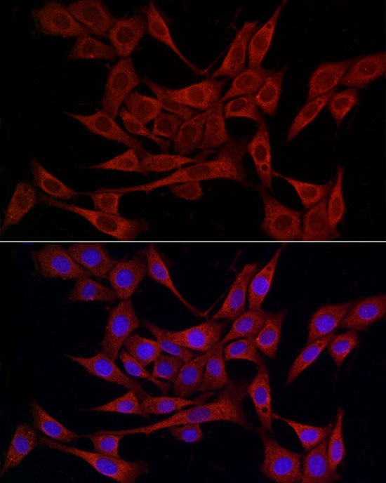
Immunofluorescence analysis of NIH/3T3 cells using PDIA4 Rabbit pAb (CAB4326) at dilution of 1:50 (40x lens). Secondary antibody: Cy3-conjugated Goat anti-Rabbit IgG (H+L) (CABS007) at 1:500 dilution. Blue: DAPI for nuclear staining.
Immunofluorescence analysis of PC-3 cells using PDIA4 Rabbit pAb (CAB4326) at dilution of 1:50 (40x lens). Secondary antibody: Cy3-conjugated Goat anti-Rabbit IgG (H+L) (CABS007) at 1:500 dilution. Blue: DAPI for nuclear staining.