The PON1 Monoclonal Antibody (CAB3441) is a high-quality antibody developed for reliable detection and analysis of target proteins. This antibody, generated in rabbits, exhibits high reactivity with human samples and is validated for use in various applications including Western blotting and immunohistochemistry.PON1, also known as Paraoxonase 1, is an enzyme primarily associated with protecting against oxidative stress and inflammation by breaking down harmful lipid peroxides. Its role in regulating lipid metabolism and preventing the formation of atherosclerotic plaques makes it a key player in cardiovascular health and disease.
This antibody is validated for use in WB, IHC-P, ELISA, IF-P applications and has demonstrated reactivity against Human, Mouse, Rat samples.
Product Name:
PON1 Monoclonal Antibody
SKU:
CAB3441
Size:
20μL, 100μL
Reactivity:
Human, Mouse, Rat
Clone Number:
ARC2001
Conjugate:
Unconjugated
Immunogen:
Synthetic peptide. This information is considered to be commercially sensitive.
Recommended starting concentration is 1 μg/mL. Please optimize the concentration based on your specific assay requirements.
Synonyms:
ESA, PON, MVCD5, PON1
Positive Sample:
Mouse liver, HepG2
Cellular Localization:
Secreted, Extracellular Space.
Calculated MW:
40kDa
Observed MW:
40kDa
This gene encodes a member of the paraoxonase family of enzymes and exhibits lactonase and ester hydrolase activity. Following synthesis in the kidney and liver, the enzyme is secreted into the circulation, where it binds to high density lipoprotein (HDL) particles and hydrolyzes thiolactones and xenobiotics, including paraoxon, a metabolite of the insecticide parathion. Polymorphisms in this gene may be associated with coronary artery disease and diabetic retinopathy. The gene is found in a cluster of three related paraoxonase genes on chromosome 7.
Purification Method
Affinity purification
Gene ID
5444
RRID
AB_2863059
Buffer Information
Store at -20℃. Avoid freeze / thaw cycles. Buffer: PBS containing 50% glycerol and 0.05% BSA, preserved with proclin300 or sodium azide, pH 7.3.
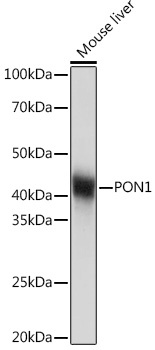
Western blot analysis of lysates from Mouse liver, using PON1 Rabbit mAb (CAB3441) at 1:1000 dilution. Secondary antibody: HRP-conjugated Goat anti-Rabbit IgG (H+L) (CABS014) at 1:10000 dilution. Lysates/proteins: 25μg per lane. Blocking buffer: 3% nonfat dry milk in TBST. Detection: ECL Basic Kit (AbGn00020). Exposure time: 1s.
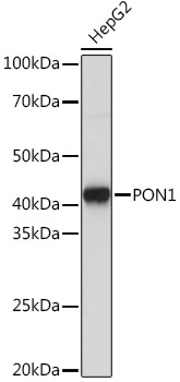
Western blot analysis of lysates from HepG2 cells, using PON1 Rabbit mAb (CAB3441) at 1:1000 dilution. Secondary antibody: HRP-conjugated Goat anti-Rabbit IgG (H+L) (CABS014) at 1:10000 dilution. Lysates/proteins: 25μg per lane. Blocking buffer: 3% nonfat dry milk in TBST. Detection: ECL Basic Kit (AbGn00020). Exposure time: 3s.
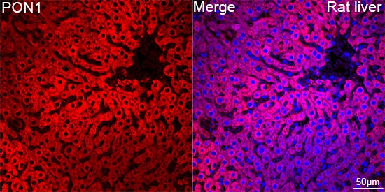
Confocal imaging of paraffin-embedded Rat liver tissue using PON1 Rabbit mAb (CAB3441, dilution 1:200) followed by a further incubation with Cy3 Goat Anti-Rabbit IgG (H+L) (CABS007, dilution 1:500) (Red). DAPI was used for nuclear staining (Blue). Objective: 40x. Perform high pressure antigen retrieval with 0.01 M citrate buffer (pH 6.0) prior to IF staining.
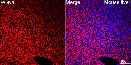
Confocal imaging of paraffin-embedded Mouse liver tissue using PON1 Rabbit mAb (CAB3441, dilution 1:200) followed by a further incubation with Cy3 Goat Anti-Rabbit IgG (H+L) (CABS007, dilution 1:500) (Red). DAPI was used for nuclear staining (Blue). Objective: 40x. Perform high pressure antigen retrieval with 0.01 M citrate buffer (pH 6.0) prior to IF staining.