The APLF Antibody (CAB17842) is a high-quality antibody developed for reliable detection and analysis of target proteins. This antibody, raised in rabbits, is highly reactive with human samples and has been validated for use in Western blot applications. The APLF Polyclonal Antibody binds specifically to the APLF protein, allowing for the detection and analysis of this important molecule in various cell types.APLF (Aprataxin and PNK-like factor) is a key player in the DNA damage response pathway, specifically in the repair of DNA double-strand breaks. Dysfunction of APLF has been linked to various diseases, including cancer and neurological disorders, highlighting its importance in maintaining cellular integrity.
This antibody is validated for use in WB, ELISA applications and has demonstrated reactivity against Rat samples.
Product Name:
APLF Antibody
SKU:
CAB17842
Size:
20μL, 100μL
Reactivity:
Rat
Conjugate:
Unconjugated
Immunogen:
Recombinant protein (or fragment).This information is considered to be commercially sensitive.
Recommended starting concentration is 1 μg/mL. Please optimize the concentration based on your specific assay requirements.
Synonyms:
APFL, PALF, Xip1, ZCCHH1, C2orf13, APLF
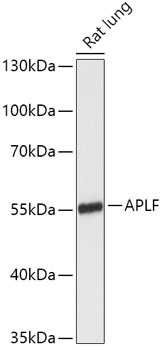
Positive Sample:
Rat lung
Cellular Localization:
Cytosol, Nucleoplasm, Nucleus.
Calculated MW:
57kDa
Observed MW:
57kDa
Enables DNA-(apurinic or apyrimidinic site) endonuclease activity; nuclease activity; and nucleotide binding activity. Involved in double-strand break repair via nonhomologous end joining and single strand break repair. Acts upstream of or within positive regulation of DNA ligation. Located in nucleoplasm. Is active in site of double-strand break.
Purification Method
Affinity purification
Gene ID
200558
RRID
AB_2768392
Buffer Information
Store at -20℃. Avoid freeze / thaw cycles. Buffer: PBS with 0.01% thimerosal,50% glycerol,pH7.3.
Western blot analysis of lysates from Rat lung, using APLF Rabbit pAb (CAB17842) at 1:1000 dilution. Secondary antibody: HRP-conjugated Goat anti-Rabbit IgG (H+L) (CABS014) at 1:10000 dilution. Lysates/proteins: 25μg per lane. Blocking buffer: 3% nonfat dry milk in TBST. Detection: ECL Enhanced Kit (AbGn00021). Exposure time: 3min.