The BCKDHB Antibody (CAB10533) is a high-quality antibody developed for reliable detection and analysis of target proteins. This complex plays a crucial role in the breakdown of branched-chain amino acids, which are essential for protein synthesis and energy production in the body.Raised in rabbits, this antibody is highly specific and reactive with human samples, making it suitable for use in Western blot applications. By binding to the BCKDHB protein, researchers can detect and analyze its expression in various cell types, providing insights into its functions and potential implications in metabolic disorders and related diseases.
This antibody is validated for use in WB, ELISA applications and has demonstrated reactivity against Mouse, Rat samples.
Product Name:
BCKDHB Antibody
SKU:
CAB10533
Size:
20μL, 100μL
Reactivity:
Mouse, Rat
Conjugate:
Unconjugated
Immunogen:
Recombinant protein (or fragment).This information is considered to be commercially sensitive.
Recommended starting concentration is 1 μg/mL. Please optimize the concentration based on your specific assay requirements.
Synonyms:
E1B, BCKDE1B, BCKDH E1-beta, BCKDHB
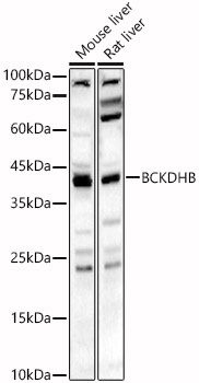
Positive Sample:
Mouse liver, Rat liver
Cellular Localization:
Mitochondrion Matrix.
Calculated MW:
43kDa
Observed MW:
43kDa
This gene encodes the E1 beta subunit of branched-chain keto acid dehydrogenase, which is a multienzyme complex associated with the inner membrane of mitochondria. This enzyme complex functions in the catabolism of branched-chain amino acids. Mutations in this gene have been associated with maple syrup urine disease (MSUD), type 1B, a disease characterized by a maple syrup odor to the urine in addition to mental and physical retardation and feeding problems. Alternative splicing at this locus results in multiple transcript variants.
Purification Method
Affinity purification
Gene ID
594
RRID
AB_2758073
Buffer Information
Store at -20℃. Avoid freeze / thaw cycles. Buffer: PBS containing 50% glycerol, preserved with proclin300 or sodium azide, pH 7.3.
Western blot analysis of various lysates, using BCKDHB Rabbit pAb (CAB10533) at 1:2000 dilution. Secondary antibody: HRP-conjugated Goat anti-Rabbit IgG (H+L) (CABS014) at 1:10000 dilution. Lysates/proteins: 25μg per lane. Blocking buffer: 3% nonfat dry milk in TBST. Detection: ECL Basic Kit (AbGn00020). Exposure time: 60s.