The Bid Antibody (CAB0210) is a high-quality antibody developed for reliable detection and analysis of target proteins. This antibody is raised in rabbits and has been validated for use in Western blot applications, demonstrating high reactivity with human samples. By binding to the BID protein, this antibody enables researchers to detect and analyze BID expression in a variety of cell types, making it an ideal tool for studies in cell biology, cancer research, and apoptosis regulation.BID, a member of the Bcl-2 protein family, functions as a critical mediator in the apoptotic pathway, linking extrinsic death receptor signaling to the intrinsic mitochondrial pathway.
This antibody is validated for use in WB, IF/ICC, ELISA applications and has demonstrated reactivity against Human, Mouse, Rat samples.
Product Name:
Bid Antibody
SKU:
CAB0210
Size:
20μL, 100μL
Reactivity:
Human, Mouse, Rat
Conjugate:
Unconjugated
Immunogen:
Recombinant protein (or fragment).This information is considered to be commercially sensitive.
This gene encodes a death agonist that heterodimerizes with either agonist BAX or antagonist BCL2, and thus regulate apoptosis. The encoded protein is a member of the BCL-2 family of cell death regulators. It is a mediator of mitochondrial damage induced by caspase-8 (CASP8); CASP8 cleaves this encoded protein, and the COOH-terminal part translocates to mitochondria where it triggers cytochrome c release. Multiple alternatively spliced transcript variants have been found.
Purification Method
Affinity purification
Gene ID
637
RRID
AB_2757024
Buffer Information
Store at -20℃. Avoid freeze / thaw cycles. Buffer: PBS containing 50% glycerol, preserved with proclin300 or sodium azide, pH 7.3.
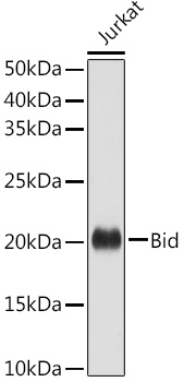
Western blot analysis of lysates from Jurkat cells, using Bid Rabbit pAb (CAB0210) at 1:1000 dilution. Secondary antibody: HRP-conjugated Goat anti-Rabbit IgG (H+L) (CABS014) at 1:10000 dilution. Lysates/proteins: 25μg per lane. Blocking buffer: 3% nonfat dry milk in TBST. Detection: ECL Basic Kit (AbGn00020). Exposure time: 10s.
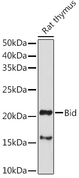
Western blot analysis of lysates from Rat thymus, using Bid Rabbit pAb (CAB0210) at 1:1000 dilution. Secondary antibody: HRP-conjugated Goat anti-Rabbit IgG (H+L) (CABS014) at 1:10000 dilution. Lysates/proteins: 25μg per lane. Blocking buffer: 3% nonfat dry milk in TBST. Detection: ECL Enhanced Kit (AbGn00021). Exposure time: 180s.