
Mycoplasma in Cell Culture Detection Guide
As a PhD student delving deep into the realm of cellular biology, there is one antagonist that consistently challenges my research: mycoplasma. This microscopic bacterium, with its ability to invisibly infiltrate and undermine cell cultures, has become a persistent foe. However, understanding the intricacies of this bacterial contaminant is the first step towards effective mitigation.
Schematic of Mycoplasma
The Invisible Enemy: Understanding Mycoplasma Contamination in Cell Culture
To grasp the significance of this issue, it's important to ask: "Why is mycoplasma bad for cell culture?". Mycoplasma is a genus of bacteria that lack a cell wall, rendering them unaffected by many common antibiotics. These small, stubborn bacteria can drastically alter cell function, morphology, metabolism, and growth characteristics. This can lead to inaccurate research data, compromised studies, and potential publication retractions if they go undetected. Moreover, their wall-less characteristic makes them resistant to common antibiotics, like penicillin, exacerbating the challenge of dealing with their contamination.
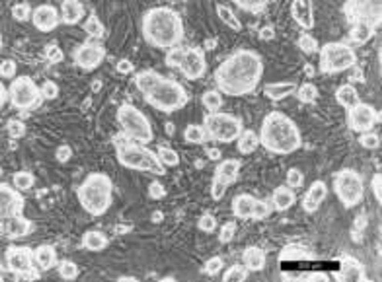
Mycoplasma
Infiltration and Propagation: How Mycoplasma Enters and Spreads in Cell Culture
The methods of mycoplasma introduction and propagation in cell culture are numerous and often insidious. In most cases, human operators are unwittingly responsible for introducing these bacteria into the sterile environment. This can happen due to improper handling techniques or contamination from the researcher’s skin or breath. Alternatively, contaminated culture media, reagents, or infected cell lines can serve as sources of mycoplasma.
Mycoplasma's modus operandi in cell culture propagation amplifies its threat. A single contaminated flask can quickly lead to a laboratory-wide problem if not identified and dealt with promptly. The bacteria can spread via aerosols generated during routine cell culture procedures, through contaminated culture media, or via shared lab equipment that hasn't been adequately sterilized.
The Art of Detection: Identifying Mycoplasma Contamination in Cell Culture
Detecting mycoplasma contamination in cell cultures can be challenging due to their ability to exist without dramatically affecting the appearance of the culture. Several detection methods are available, including DNA staining, enzyme activity, PCR assays, and culture isolation.
For instance, using DAPI (4',6-diamidino-2-phenylindole), a fluorescent stain that binds strongly to A-T rich regions in DNA, can be employed. Under UV light, DAPI-stained DNA exhibits a bright blue fluorescence, allowing visualization of potential mycoplasma contamination.
Furthermore, PCR-based detection assays are considered the gold standard due to their high sensitivity and specificity. These tests amplify the DNA of the contaminant, allowing for easier detection even at low concentrations.

Managing the Menace: Treating and Avoiding Mycoplasma Contamination
Once a culture has been identified as contaminated, immediate action is necessary. Many researchers ask, "How to get rid of mycoplasma contamination in cell culture?" The standard answer, although painful, is to discard the contaminated cultures entirely. If this is not an option, a number of commercial kits are available that contain antibiotics such as tetracyclines or fluoroquinolones, which can be effective against mycoplasma.
However, prevention is better than cure. It is critical to maintain stringent aseptic techniques during all stages of cell culture. Regular testing of cell cultures and the use of mycoplasma-free media and reagents also contribute to a clean cell culture environment.
The Complex Task of Culturing Mycoplasma
Though not routinely performed in cell culture labs, mycoplasma can be cultured under specific conditions. Mycoplasma pneumoniae, for example, can grow on Eaton’s agar, but it requires precise conditions and a very specific environment, including sterols and a protein source.
To conclude, the battle against mycoplasma contamination is a persistent challenge in cell culture labs. Understanding the nature of these bacteria, maintaining rigorous cell culture practices, and deploying effective detection strategies are key to managing their impact. While it may seem a daunting task, successful containment of mycoplasma is not only feasible but critical to ensuring the accuracy and reliability of your cell culture research.
Related Resources
Written by Sean Mac Fhearraigh
Seán Mac Fhearraigh PhD is a co-founder of Assay Genie. Seán carried out his undergraduate degree in Genetics at Trinity College Dublin, followed by a PhD at University College Dublin. He carried out a post-doc at the Department of Genetics, University of Cambridge. Seán is now Chief Technical Officer at Assay Genie.
Recent Posts
-
ELISA vs Western Blot: Which Technique Should You Choose?
Written by Seán Mac Fhearraigh, PhD • Updated: 19 May 2026 • ~9 min read Quick Answer ELISA …20th May 2026 -
Types of ELISA: Direct, Indirect, Sandwich & Competitive Compared
Written by Seán Mac Fhearraigh, PhD • Updated: 19 May 2026 • ~9 min read Quick Answer The fi …20th May 2026 -
Sandwich ELISA: Step-by-Step Protocol & Troubleshooting
Written by Seán Mac Fhearraigh, PhD • Updated: 19 May 2026 • ~ …20th May 2026