
Serum vs Plasma: Key Differences & When to Use Each [Guide]
Serum vs Plasma: Key Differences and When to Use Each
Serum and plasma come from the same blood draw but are not interchangeable. One is collected after clotting, the other before it, and that single difference changes which analytes survive, which are artefactually raised, and which assays will give you a trustworthy number. This guide covers the composition difference, collection protocols for both, tube selection, and how to choose the right matrix for an ELISA.
Browse ELISA kits →Key takeaways
- Plasma is obtained from anticoagulated blood and retains clotting factors; serum is what remains after blood has clotted, so those factors are consumed and absent.
- Fibrinogen and the coagulation factors can only be measured in plasma — they are gone from serum by definition.
- Platelet-derived analytes are artefactually elevated in serum, because clotting activates platelets and releases their contents.
- Serum is generally preferred for enzyme measurements, since anticoagulants such as EDTA chelate the metal ions some enzymes require.
- Plasma is not one matrix: EDTA, heparin and citrate plasma behave differently, and an assay validated in one is not automatically valid in another.
- Plasma gives a slightly higher yield per draw and can be processed immediately, which matters when sample volume or turnaround is limited.
- Always check the validated matrix on the kit datasheet, and never mix matrices within a single experiment.
ELISA kits where the matrix matters
Each kit below illustrates a different aspect of the serum-versus-plasma decision — analytes that exist only in plasma, analytes corrupted by clotting, and analytes where serum is the better choice.
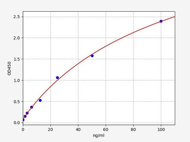
Human Fibrinogen ELISA Kit
Plasma only — fibrinogen is converted to fibrin during clotting and is absent from serum.
View kit →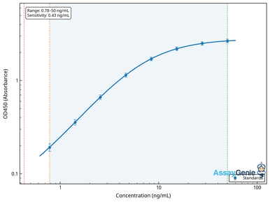
Human Prothrombin / Factor II ELISA Kit
A coagulation factor consumed in the clotting cascade, so it requires anticoagulated plasma.
View kit →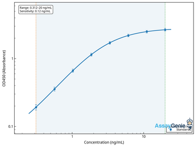
Human Platelet Factor 4 ELISA Kit
Released from activated platelets — grossly elevated in serum, so plasma is essential.
View kit →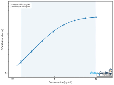
Human Alanine Aminotransferase ELISA Kit
Serum is generally preferred for enzymes, since chelating anticoagulants can affect activity.
View kit →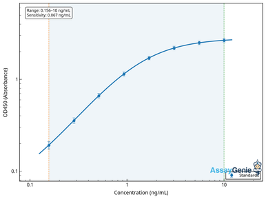
Human C-Reactive Protein ELISA Kit
An acute-phase protein measurable in either matrix — but keep the choice consistent across a study.
View kit →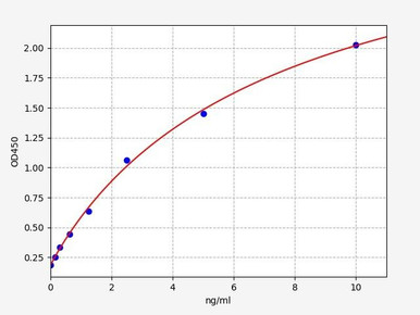
Human Insulin ELISA Kit
Sensitive to handling delays; matrix and processing time both affect the result.
View kit →What are serum and plasma?
Whole blood is a suspension: red cells, white cells and platelets carried in a straw-coloured liquid. Separating that liquid from the cells is straightforward, but how you separate it determines what you end up with.
Plasma is the liquid fraction recovered when blood is prevented from clotting. An anticoagulant is added at the moment of collection, the sample is centrifuged, and the plasma sits above the buffy coat and packed red cells. Because clotting never occurred, plasma still contains fibrinogen and the full set of coagulation factors.
Serum is what remains after blood has been allowed to clot. No anticoagulant is used; the sample is left to coagulate, the clot is removed by centrifugation, and the clear fluid above it is serum. The clot has taken the fibrinogen with it — converted to fibrin — along with the other factors consumed in the cascade.
How their composition differs
Serum and plasma share the great majority of their contents: water, electrolytes, glucose and other nutrients, hormones, dissolved gases, waste products and the bulk of plasma proteins including albumin and the immunoglobulins. The differences are narrow but consequential.
The clearest is fibrinogen and the coagulation factors, present in plasma and absent from serum. The second, less obvious and more often responsible for confusing results, is the platelet releasate. Clotting activates platelets, which degranulate and discharge their contents into the surrounding fluid. Anything stored in platelet granules is therefore substantially higher in serum than in plasma — not because the patient had more of it, but because the tube manufactured it. Serotonin, platelet factor 4, and several growth factors behave this way.
Two further differences are worth noting. Serum has a marginally higher total protein concentration once fibrinogen is removed and water redistributes, and plasma is diluted very slightly by the anticoagulant volume in the tube. Neither matters for most assays, but both argue against comparing absolute values obtained in different matrices.
How to collect serum
Serum collection is simple but time-dependent: the clot must form fully before centrifugation, or fibrin will continue forming in the separated sample and interfere with downstream assays.
Materials required
- Plain collection tubes with no anticoagulant — red or gold top
- Sterile needle and syringe, or a vacutainer system
- Centrifuge, ideally refrigerated
- Pipettes and labelled storage tubes
- Personal protective equipment: gloves, lab coat, eye protection
| Step | Procedure |
|---|---|
| 1 | Put on personal protective equipment. |
| 2 | Clean the venipuncture site with an appropriate antiseptic. |
| 3 | Perform venipuncture and collect blood into a plain tube containing no anticoagulant. |
| 4 | Leave the tube undisturbed at room temperature for 15 to 30 minutes to allow the clot to form completely. |
| 5 | Centrifuge at 1,000 to 2,000 ×g for 10 minutes, preferably refrigerated. |
| 6 | Pipette the serum — the clear upper layer — into a fresh tube without disturbing the clot. |
| 7 | Label as serum, and freeze promptly if not assayed the same day. |
Do not shorten the clotting step to save time. Incompletely clotted samples are the most common cause of fibrin strands appearing in thawed serum, which block pipette tips and scatter light in plate readers.
How to collect plasma
Plasma can be separated immediately, which is its practical advantage, but it demands correct mixing at the point of collection and prompt centrifugation.
| Step | Procedure |
|---|---|
| 1 | Put on personal protective equipment. |
| 2 | Clean the venipuncture site with an appropriate antiseptic. |
| 3 | Perform venipuncture and collect blood into a tube containing the required anticoagulant. |
| 4 | Invert the tube gently 5 to 10 times immediately, to mix anticoagulant through the sample and prevent clotting. Do not shake. |
| 5 | Fill the tube to its stated volume, so the anticoagulant-to-blood ratio is correct. |
| 6 | Centrifuge within two hours of the draw, or sooner if the assay requires it. |
| 7 | Pipette the plasma without disturbing the buffy coat or red cells beneath it. |
| 8 | Label as plasma and record which anticoagulant was used. |
That last step matters more than it looks. Recording the anticoagulant is not administrative tidiness — EDTA, heparin and citrate plasma are chemically different matrices, and a sample labelled only “plasma” may be unusable for an assay validated in one specific type.
Which tube produces which matrix
Tube cap colour indicates the additive. Conventions vary slightly between manufacturers and regions, so confirm against your supplier, but the common scheme is below.
| Cap colour | Additive | Yields | Typical use |
|---|---|---|---|
| Red or gold (tiger) | None, or clot activator and gel | Serum | Biochemistry, enzymes, serology, most ELISA work |
| Lavender or purple | EDTA | Plasma | Haematology; the most common plasma type for immunoassay |
| Green | Heparin | Plasma | Some clinical chemistry and cell-based work |
| Light blue | Sodium citrate | Plasma | Coagulation testing, where the ratio to blood is critical |
| Grey | Fluoride and oxalate | Plasma | Glucose and lactate, where glycolysis must be halted |
Choosing between serum and plasma
In practice the decision is usually made for you by the analyte or the kit. Where you do have a choice, these are the considerations that matter.
Choose plasma when the target is a coagulation factor or fibrinogen, when the analyte is stored in platelets and would be released by clotting, when you need the sample separated immediately, or when volume is tight — plasma yields roughly 15 to 20 per cent more liquid from the same draw, because no volume is lost to the clot.
Choose serum when measuring enzymes whose activity depends on metal ions that chelating anticoagulants would sequester, when an assay was validated in serum, or when you want to avoid introducing an anticoagulant that might interfere with antibody binding. Serum is also the conventional matrix for serology.
Whichever you pick, the overriding rule is consistency. Absolute concentrations are not reliably comparable between matrices, so a study that mixes serum and plasma samples has introduced a variable that no amount of statistics will remove afterwards.
Analytes that dictate the matrix
Plasma-only analytes
The coagulation cascade is consumed during clotting, so its components cannot be recovered from serum. Fibrinogen, prothrombin (factor II), and factors V, VII, VIII, IX, X, XI, XII and XIII all require anticoagulated plasma. Citrate plasma is the reference matrix for functional coagulation testing.
Analytes corrupted by clotting
Platelet contents are released during clot formation, so serum values reflect the tube rather than the circulation. Platelet factor 4, serotonin, thromboxane and platelet-derived growth factors are all affected. If a platelet-associated analyte is your endpoint, plasma is not merely preferable but necessary — and even then, careful centrifugation matters, since platelet carryover into plasma produces the same artefact in miniature.
Analytes where serum is preferred
Enzyme measurements are generally better in serum. Several enzymes depend on divalent cations, and EDTA chelates them, which can suppress apparent activity. Alanine aminotransferase, aspartate aminotransferase, alkaline phosphatase, gamma-glutamyl transferase, lactate dehydrogenase and creatine kinase are conventionally measured in serum for this reason.
Analytes measurable in either
Many soluble proteins, hormones and cytokines can be measured in both, including C-reactive protein, albumin, the immunoglobulins and most interleukins. Even here the two matrices may give slightly different absolute values, so pick one at the design stage and stay with it.
Serum and plasma in ELISA
ELISA quantifies a target by capturing it on an immobilised antibody and detecting it with a second, enzyme-linked antibody, generating a colour change proportional to concentration. Both serum and plasma are routine matrices, and most commercial kits are validated for one or both.
What makes matrix choice consequential in ELISA is that blood-derived samples are complex, and their non-target components affect the assay. Fibrinogen in plasma adds viscosity and can raise background. Residual fibrin in under-clotted serum scatters light and blocks tips. Heparin can interfere with some antibody-antigen interactions. EDTA can affect assays that depend on divalent cations. None of this makes either matrix wrong — it means the validated matrix on the datasheet is a real constraint rather than a suggestion.
Two practical rules follow. Check the validated matrix and the stated dynamic range before designing the experiment, and if you must switch matrix mid-project, treat it as a change requiring re-validation and a fresh standard curve rather than a substitution.
Pre-analytical pitfalls
Most unexplained immunoassay results trace back to how the sample was handled rather than to the kit. The recurring culprits:
- Haemolysis. Ruptured red cells release haemoglobin and intracellular contents, discolouring the sample and interfering optically and chemically. Usually caused by a needle that is too fine, excessive suction, or vigorous shaking.
- Delayed separation. Cells continue to metabolise while in contact with the liquid fraction. Glucose falls, potassium and lactate dehydrogenase rise. Separate plasma within two hours.
- Incomplete clotting. Spinning serum too early leaves fibrinogen to polymerise later, producing strands in the thawed sample.
- Platelet carryover. Under-centrifuged plasma retains platelets that release their contents on freezing and thawing, mimicking a serum artefact.
- Repeated freeze-thaw. Each cycle degrades labile analytes and generates precipitates. Aliquot before freezing so each sample is thawed once.
- Lipaemia. Turbid, lipid-rich samples from non-fasting subjects scatter light and can shift readings.
- Wrong fill volume. An underfilled anticoagulant tube over-dilutes the sample and distorts the anticoagulant ratio, which matters most for citrate.
References
- Lippi G, Plebani M. Serum versus plasma: which is the better choice? Clinical Chemistry and Laboratory Medicine, 2012;50(9):1523–1524.
- Van Der Meer PF, Pietersz RNI. Blood plasma and serum components. Wiley Encyclopedia of Blood Transfusion, 2016.
- Clinical and Laboratory Standards Institute guidance on the collection and handling of diagnostic blood specimens remains the reference standard for pre-analytical procedure.
Choosing an ELISA kit
Human, mouse and rat ELISA kits with the validated sample matrix and dynamic range stated on every datasheet — so you can confirm serum or plasma compatibility before you commit samples.
Browse ELISA kits →Frequently asked questions
What is the main difference between serum and plasma?
Plasma is collected from blood treated with an anticoagulant, so it still contains fibrinogen and the coagulation factors. Serum is collected after blood has clotted, so those factors have been consumed and are absent. Everything else is largely shared.
Can I use serum instead of plasma for my ELISA?
Only if the kit is validated for serum. Some analytes cannot be measured in serum at all — fibrinogen and coagulation factors are consumed by clotting — and platelet-derived analytes are artefactually elevated in serum. Check the datasheet before substituting.
Which gives more sample, serum or plasma?
Plasma, by roughly 15 to 20 per cent from the same draw, because no volume is trapped in the clot. Plasma can also be separated immediately rather than waiting 15 to 30 minutes for clotting.
Why is serum preferred for enzyme assays?
Because chelating anticoagulants such as EDTA bind divalent cations, and several enzymes require those ions for activity. Measuring them in EDTA plasma can therefore understate activity. Serum avoids the problem entirely.
Are EDTA, heparin and citrate plasma interchangeable?
No. They are chemically distinct matrices and can affect assays differently — heparin may interfere with some antibody binding, EDTA affects cation-dependent reactions, and citrate dilutes the sample by a fixed ratio. An assay validated in one is not automatically valid in another.
How quickly must blood be processed?
Separate plasma within about two hours of the draw, since cells continue metabolising and will alter glucose, potassium and lactate dehydrogenase. For serum, allow 15 to 30 minutes for full clotting first, then centrifuge promptly.
Why does my thawed serum contain stringy material?
Almost always incomplete clotting before centrifugation. Fibrinogen left in the separated serum polymerises into fibrin strands later. Allowing the full clotting time prevents it.
Seán Mac Fhearraigh is a co-founder of Assay Genie. He completed his undergraduate degree in Genetics at Trinity College Dublin, followed by a PhD at University College Dublin, and a post-doc at the Department of Genetics, University of Cambridge. Seán is Chief Technical Officer at Assay Genie.
Recent Posts
-
Competitive vs Sandwich ELISA: Should You Subtract the Blank?
Quick answer: A researcher subtracted the blank from a competitive ELISA (a Serotonin ki …21st Aug 2026 -
Antigen Processing and Presentation: MHC-I, MHC-II, Cross-Priming
Every T-cell response begins with a protein being taken apart. Before a CD8 or CD4 T cell can see a …20th Aug 2026 -
Apoptosis Pathway: Death Receptor, Mitochondrial and Granzyme Routes
Apoptosis is not one pathway but three routes into the same execution machinery. A death ligand at …20th Aug 2026