The CTSG Antibody (CAB13172) is a high-quality antibody developed for reliable detection and analysis of target proteins. This antibody, generated in rabbits, is highly specific to human samples and is validated for Western blot applications. By binding to CTSG, the antibody enables accurate detection and analysis of this protein in various cell types, making it ideal for research in immunology, inflammation, and diseases like cancer.CTSG is known for its role in immune cell function, particularly in neutrophils where it participates in processes like antimicrobial defense and inflammation regulation.
This antibody is validated for use in WB, IHC-P, ELISA applications and has demonstrated reactivity against Human, Mouse, Rat samples.
Product Name:
CTSG Antibody
SKU:
CAB13172
Size:
20μL, 100μL
Reactivity:
Human, Mouse, Rat
Conjugate:
Unconjugated
Immunogen:
Recombinant protein (or fragment).This information is considered to be commercially sensitive.
Recommended starting concentration is 1 μg/mL. Please optimize the concentration based on your specific assay requirements.
Synonyms:
CG, CATG, Cathepsin G
Positive Sample:
Mouse kidney, Rat kidney
Cellular Localization:
Cell Surface.
Calculated MW:
29kDa
Observed MW:
29kDa
The protein encoded by this gene, a member of the peptidase S1 protein family, is found in azurophil granules of neutrophilic polymorphonuclear leukocytes. The encoded protease has a specificity similar to that of chymotrypsin C, and may participate in the killing and digestion of engulfed pathogens, and in connective tissue remodeling at sites of inflammation. In addition, the encoded protein is antimicrobial, with bacteriocidal activity against S. aureus and N. gonorrhoeae. Transcript variants utilizing alternative polyadenylation signals exist for this gene.
Purification Method
Affinity purification
Gene ID
1511
RRID
AB_2760023
Buffer Information
Store at -20℃. Avoid freeze / thaw cycles. Buffer: PBS with 0.01% thimerosal,50% glycerol,pH7.3.
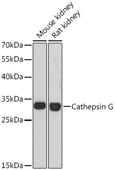
Western blot analysis of various lysates using Cathepsin G Rabbit pAb (CAB13172) at 1:3000 dilution. Secondary antibody: HRP-conjugated Goat anti-Rabbit IgG (H+L) (CABS014) at 1:10000 dilution. Lysates/proteins: 25μg per lane. Blocking buffer: 3% nonfat dry milk in TBST. Detection: ECL Basic Kit (AbGn00020). Exposure time: 90s.
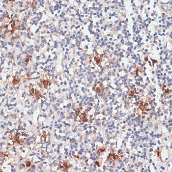
Immunohistochemistry analysis of paraffin-embedded Human tonsil using Cathepsin G Rabbit pAb (CAB13172) at dilution of 1:100 (40x lens). Microwave antigen retrieval performed with 0.01M PBS Buffer (pH 7.2) prior to IHC staining.
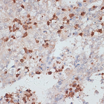
Immunohistochemistry analysis of paraffin-embedded Human uterine cancer using Cathepsin G Rabbit pAb (CAB13172) at dilution of 1:100 (40x lens). Microwave antigen retrieval performed with 0.01M PBS Buffer (pH 7.2) prior to IHC staining.
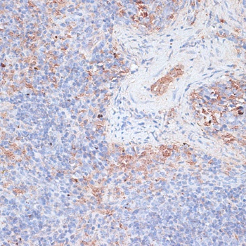
Immunohistochemistry analysis of paraffin-embedded Mouse spleen using Cathepsin G Rabbit pAb (CAB13172) at dilution of 1:100 (40x lens). Microwave antigen retrieval performed with 0.01M PBS Buffer (pH 7.2) prior to IHC staining.