The CD3D Antibody (CAB1238) is a high-quality antibody developed for reliable detection and analysis of target proteins. This polyclonal antibody is raised in rabbits and has been validated for use in Western blot applications, showing high reactivity with human samples. By specifically binding to the CD3D protein, this antibody enables accurate detection and analysis in various cell types, making it ideal for studies in immunology, infectious diseases, and cancer research.CD3D is essential for T-cell activation and function, playing a crucial role in the adaptive immune response. Dysregulation of CD3D expression or function has been implicated in various diseases, including autoimmune disorders, immunodeficiency conditions, and certain types of cancer.
This antibody is validated for use in WB, IHC-P, IF/ICC, ELISA applications and has demonstrated reactivity against Human, Rat samples.
Product Name:
CD3D Antibody
SKU:
CAB1238
Size:
20μL, 100μL
Reactivity:
Human, Rat
Conjugate:
Unconjugated
Immunogen:
Recombinant protein (or fragment).This information is considered to be commercially sensitive.
Recommended starting concentration is 1 μg/mL. Please optimize the concentration based on your specific assay requirements.
Synonyms:
T3D, IMD19, CD3DELTA, CD3-DELTA, CD3D
Positive Sample:
Jurkat
Cellular Localization:
Membrane, Single-Pass Type I Membrane Protein.
Calculated MW:
19kDa
Observed MW:
19kDa
The protein encoded by this gene is part of the T-cell receptor/CD3 complex (TCR/CD3 complex) and is involved in T-cell development and signal transduction. The encoded membrane protein represents the delta subunit of the CD3 complex, and along with four other CD3 subunits, binds either TCR alpha/beta or TCR gamma/delta to form the TCR/CD3 complex on the surface of T-cells. Defects in this gene are a cause of severe combined immunodeficiency autosomal recessive T-cell-negative/B-cell-positive/NK-cell-positive (SCIDBNK). Two transcript variants encoding different isoforms have been found for this gene. Other variants may also exist, but the full-length natures of their transcripts has yet to be defined.
Purification Method
Affinity purification
Gene ID
915
RRID
AB_2759223
Buffer Information
Store at -20℃. Avoid freeze / thaw cycles. Buffer: PBS containing 50% glycerol, preserved with proclin300 or sodium azide, pH 7.3.
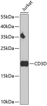
Western blot analysis of lysates from Jurkat cells, using CD3D Rabbit pAb (CAB1238) at 1:500 dilution._Secondary antibody: HRP-conjugated Goat anti-Rabbit IgG (H+L) (CABS014) at 1:10000 dilution._Lysates/proteins: 25μg per lane._Blocking buffer: 3% nonfat dry milk in TBST._Detection: ECL Enhanced Kit (AbGn00021)._Exposure time: 90s.
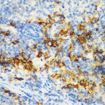
Immunohistochemistry analysis of paraffin-embedded Rat spleen using CD3D Rabbit pAb (CAB1238) at dilution of 1:200 (40x lens). Microwave antigen retrieval performed with 0.01M PBS Buffer (pH 7.2) prior to IHC staining.