The CFD Monoclonal Antibody (CAB21110) is a high-quality antibody developed for reliable detection and analysis of target proteins. This antibody, produced through hybridoma technology, is highly specific to CFD and has been validated for use in various applications such as ELISA and immunofluorescence.CFD, also known as complement factor D, plays a crucial role in the alternative pathway of the complement system by cleaving complement component C3. Dysregulation of the complement system has been linked to various diseases including autoimmune disorders, inflammatory conditions, and infectious diseases.
This antibody is validated for use in WB, ELISA applications and has demonstrated reactivity against Human samples.
Product Name:
CFD Monoclonal Antibody
SKU:
CAB21110
Size:
20μL, 100μL
Reactivity:
Human
Clone Number:
ARC2997
Conjugate:
Unconjugated
Immunogen:
Recombinant protein (or fragment).This information is considered to be commercially sensitive.
Recommended starting concentration is 1 μg/mL. Please optimize the concentration based on your specific assay requirements.
Synonyms:
DF, ADN, PFD, ADIPSIN, CFD
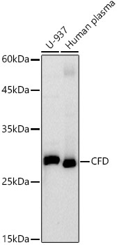
Positive Sample:
U-937, Human plasma
Cellular Localization:
Secreted.
Calculated MW:
27kDa
Observed MW:
27kDa
This gene encodes a member of the S1, or chymotrypsin, family of serine peptidases. This protease catalyzes the cleavage of factor B, the rate-limiting step of the alternative pathway of complement activation. This protein also functions as an adipokine, a cell signaling protein secreted by adipocytes, which regulates insulin secretion in mice. Mutations in this gene underlie complement factor D deficiency, which is associated with recurrent bacterial meningitis infections in human patients. Alternative splicing of this gene results in multiple transcript variants. At least one of these variants encodes a preproprotein that is proteolytically processed to generate the mature protease.
Purification Method
Affinity purification
Gene ID
1675
Buffer Information
Store at -20℃. Avoid freeze / thaw cycles. Buffer: PBS containing 50% glycerol and 0.05% BSA, preserved with proclin300 or sodium azide, pH 7.3.
Western blot analysis of various lysates using CFD Rabbit mAb (CAB21110) at1:1000 dilution. Secondary antibody: HRP-conjugated Goat anti-Rabbit IgG (H+L) (CABS014) at 1:10000 dilution. Lysates/proteins: 25μg per lane. Blocking buffer: 3% nonfat dry milk in TBST. Detection: ECL Basic Kit (AbGn00020). Exposure time: 90s.
 at1:1000 dilution. Secondary antibody: HRP Goat Anti-Rabbit IgG (H+L) at 1:10000 dilution. Lysates/proteins: 25μg per lane. Blocking buffer: 3% nonfat dry milk in TBST.")
 at1:1000 dilution. Secondary antibody: HRP Goat Anti-Rabbit IgG (H+L) at 1:10000 dilution. Lysates/proteins: 25μg per lane. Blocking buffer: 3% nonfat dry milk in TBST.")


")