The COPG2 Antibody (CAB15816) is a high-quality antibody developed for reliable detection and analysis of target proteins. This antibody, generated in rabbits, demonstrates high reactivity with human samples and is optimized for Western blot applications. By specifically binding to COPG2, this antibody allows for the precise detection and analysis of the protein in various cell types, making it an essential component in studies related to cell biology and molecular mechanisms.COPG2 plays a crucial role in maintaining the integrity of the Golgi apparatus and facilitating the transport of proteins within the cell.
This antibody is validated for use in WB, IHC-P, IF/ICC, ELISA applications and has demonstrated reactivity against Human, Mouse, Rat samples.
Product Name:
COPG2 Antibody
SKU:
CAB15816
Size:
20μL, 100μL
Reactivity:
Human, Mouse, Rat
Conjugate:
Unconjugated
Immunogen:
Recombinant protein (or fragment).This information is considered to be commercially sensitive.
Predicted to enable structural molecule activity. Involved in intra-Golgi vesicle-mediated transport. Part of COPI vesicle coat.
Purification Method
Affinity purification
Gene ID
26958
RRID
AB_2763239
Buffer Information
Store at -20℃. Avoid freeze / thaw cycles. Buffer: PBS with 0.01% thimerosal,50% glycerol,pH7.3.
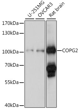
Western blot analysis of various lysates using COPG2 Rabbit pAb (CAB15816) at 1:1000 dilution. Secondary antibody: HRP-conjugated Goat anti-Rabbit IgG (H+L) (CABS014) at 1:10000 dilution. Lysates/proteins: 25μg per lane. Blocking buffer: 3% nonfat dry milk in TBST. Detection: ECL Basic Kit (AbGn00020). Exposure time: 90s.
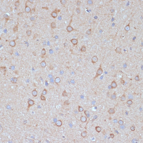
Immunohistochemistry analysis of paraffin-embedded Rat brain using COPG2 Rabbit pAb (CAB15816) at dilution of 1:100 (40x lens). Microwave antigen retrieval performed with 0.01M PBS Buffer (pH 7.2) prior to IHC staining.
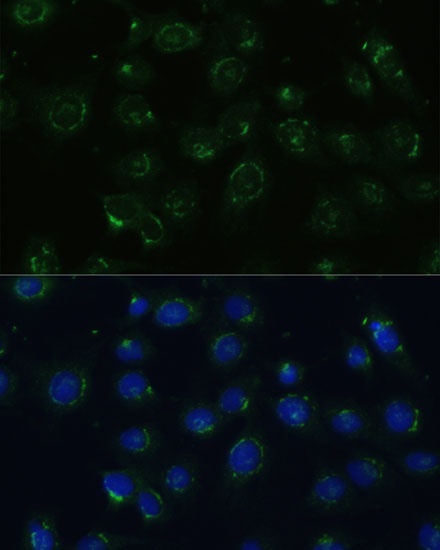
Immunofluorescence analysis of C6 cells using COPG2 Rabbit pAb (CAB15816) at dilution of 1:100 (40x lens). Secondary antibody: Cy3-conjugated Goat anti-Rabbit IgG (H+L) (CABS007) at 1:500 dilution. Blue: DAPI for nuclear staining.
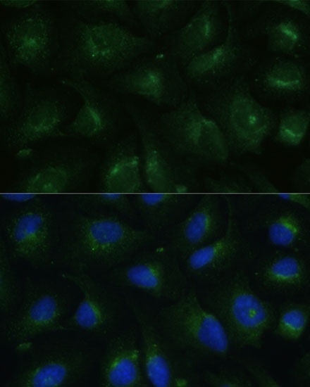
Immunofluorescence analysis of U-2 OS cells using COPG2 Rabbit pAb (CAB15816) at dilution of 1:100 (40x lens). Secondary antibody: Cy3-conjugated Goat anti-Rabbit IgG (H+L) (CABS007) at 1:500 dilution. Blue: DAPI for nuclear staining.