The TCEB2 Antibody (CAB5362) is a high-quality antibody developed for reliable detection and analysis of target proteins. This antibody, raised in rabbits, exhibits high reactivity with Elongin B in human samples and has been validated for use in Western blot applications.Elongin B is known to play a crucial role in the assembly of the Elongin BC complex, which functions as a substrate recognition module in the ubiquitin ligase complex. This complex is involved in targeting specific proteins for degradation, making Elongin B an important player in cellular processes such as gene expression and protein turnover.
This antibody is validated for use in WB, IP, ELISA applications and has demonstrated reactivity against Human, Mouse samples.
Product Name:
TCEB2 Antibody
SKU:
CAB5362
Size:
20μL, 100μL
Reactivity:
Human, Mouse
Conjugate:
Unconjugated
Immunogen:
Recombinant protein (or fragment).This information is considered to be commercially sensitive.
This gene encodes the protein elongin B, which is a subunit of the transcription factor B (SIII) complex. The SIII complex is composed of elongins A/A2, B and C. It activates elongation by RNA polymerase II by suppressing transient pausing of the polymerase at many sites within transcription units. Elongin A functions as the transcriptionally active component of the SIII complex, whereas elongins B and C are regulatory subunits. Elongin A2 is specifically expressed in the testis, and capable of forming a stable complex with elongins B and C. The von Hippel-Lindau tumor suppressor protein binds to elongins B and C, and thereby inhibits transcription elongation. Two alternatively spliced transcript variants encoding different isoforms have been described for this gene. Pseudogenes have been identified on chromosomes 11 and 13.
Purification Method
Affinity purification
Gene ID
6923
RRID
AB_2766172
Buffer Information
Store at -20℃. Avoid freeze / thaw cycles. Buffer: PBS containing 50% glycerol, preserved with proclin300 or sodium azide, pH 7.3.
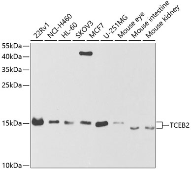
Western blot analysis of various lysates using TCEB2 Rabbit pAb (CAB5362) at 1:1000 dilution. Secondary antibody: HRP-conjugated Goat anti-Rabbit IgG (H+L) (CABS014) at 1:10000 dilution. Lysates/proteins: 25μg per lane. Blocking buffer: 3% nonfat dry milk in TBST. Detection: ECL Basic Kit (AbGn00020). Exposure time: 90s.
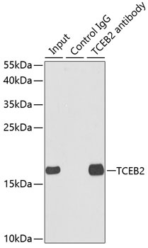
Immunoprecipitation analysis of 150 μg extracts of MCF7 cells using 3 μg TCEB2 antibody (CAB5362). Western blot was performed from the immunoprecipitate using TCEB2 antibody (CAB5362) at a dilution of 1:500.