The Erlin-2 Monoclonal Antibody (CAB0781) is a high-quality antibody developed for reliable detection and analysis of target proteins. This polyclonal antibody, raised in rabbits, is highly specific to human samples and has been validated for use in Western blot applications.Erlin-2 has been implicated in various cellular functions, including endoplasmic reticulum-associated protein degradation and lipid droplet biogenesis.
This antibody is validated for use in WB, IHC-P, ELISA applications and has demonstrated reactivity against Human, Mouse, Rat samples.
Product Name:
Erlin-2 Monoclonal Antibody
SKU:
CAB0781
Size:
20μL, 100μL
Reactivity:
Human, Mouse, Rat
Clone Number:
ARC2538
Conjugate:
Unconjugated
Immunogen:
Synthetic peptide. This information is considered to be commercially sensitive.
Recommended starting concentration is 1 μg/mL. Please optimize the concentration based on your specific assay requirements.
Synonyms:
NET32, SPFH2, SPG18, C8orf2, Erlin-2
Positive Sample:
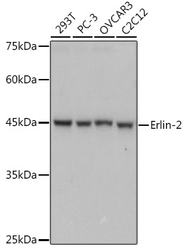
293T, PC-3, OVCAR3, C2C12
Cellular Localization:
Endoplasmic Reticulum Membrane, Single-Pass Type Ii Membrane Protein.
Calculated MW:
38kDa
Observed MW:
43kDa
This gene encodes a member of the SPFH domain-containing family of lipid raft-associated proteins. The encoded protein is localized to lipid rafts of the endoplasmic reticulum and plays a critical role in inositol 1,4,5-trisphosphate (IP3) signaling by mediating ER-associated degradation of activated IP3 receptors. Mutations in this gene are a cause of spastic paraplegia-18 (SPG18). Alternatively spliced transcript variants encoding multiple isoforms have been observed for this gene.
Purification Method
Affinity purification
Gene ID
11160
Buffer Information
Store at -20℃. Avoid freeze / thaw cycles. Buffer: PBS containing 50% glycerol and 0.05% BSA, preserved with proclin300 or sodium azide, pH 7.3.
Western blot analysis of various lysates using Erlin-2 Rabbit mAb (CAB0781) at 1:1000 dilution. Secondary antibody: HRP-conjugated Goat anti-Rabbit IgG (H+L) (CABS014) at 1:10000 dilution. Lysates/proteins: 25μg per lane. Blocking buffer: 3% nonfat dry milk in TBST. Detection: ECL Basic Kit (AbGn00020). Exposure time: 1s.
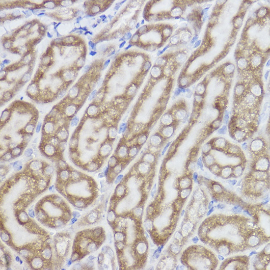
Immunohistochemistry analysis of paraffin-embedded Mouse kidney using Erlin-2 Rabbit mAb (CAB0781) at dilution of 1:100 (40x lens). High pressure antigen retrieval performed with 0.01M Citrate buffer (pH 6.0) prior to IHC staining.